Download
1 / 20

E N D
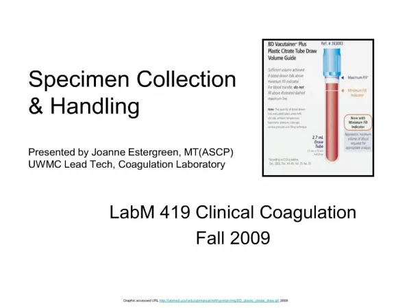
1. Specimen Collection & Handling LabM 419 Clinical Coagulation
Fall 2009
3. Doctors want to know . . . Why is my patient bleeding?
How do I stop the bleeding?
Is my treatment working to prevent further bleeding?
Why did my patient thrombose (stroke or heart attack)?
Is my treatment of the patient working to prevent further thrombosis?
4. CLOTTING ASSAYS Screen for coagulation dysfunction resulting in bleeding
Monitor anti-thrombosis treatment
Monitor transfusion therapy to treat the bleeding patient.
Add clot initiator to sample
5. Samples Collected For Coagulation Studies Maintain stability of Coagulation Factors
Prevent clot formation in sample prior to testing
Prevent Factor activation in tube - even if clot does not form
Additives in tube must not interfere with individual Coagulation Factors
6. Samples are Drawn into Tubes Tubes contain a vacuum - will fill without forcing
Coagulation Tubes - Powder Blue Top
Non-wettable, siliconized surface or plastic tubes so as not to activate factors
Contains buffered Sodium Citrate which�
prevents clotting
provides environment where clotting factors are stable for hours
when testing occurs, helps maintain body pH
Anticoagulant complexes calcium and is easily overcome by adding small amounts of calcium back until anticoagulant is overcome and the clotting endpoint can be formed
7. Drawing Good Quality Coagulation Samples No Trauma.
Smooth blood flow into tube.
Vacutainer system collects sample directly into tube.
Multiple tube draws, draw in a certain order. We recommend:
Sterile blood cultures
Discard tube (yes/no � if blue top first tube)
Blue top
Lavender top
Green top, gold top etc.
Discard Tubes being drawn prior to blue top is no longer necessary per CLSI (formerly NCCLS) if blue top drawn first and standard tests requested. Still recommended when drawing samples for specialized coagulation testing and is necessary if drawing from a line catheter.
Syringe draws are not recommended. If veins are highly collapsible then syringe may be needed. Use small volume syringe (< 20 mL).
19 - 22 gauge needles to avoid hemolysis in adults and 21 - 23 for pediatric samples. *note - larger the gauge the smaller the bore diameter of the needle.
8. STRATEGIES FOR SUCCESSFUL COAGULATION SAMPLE COLLECTION Main problem sample types
Clotted
�Short�
insufficient tube fill
Hemolyzed samples
9. Clotted Samples Clotting factors pre-activated
Clotting factors consumed in clot
Often caused by inadequate anticoagulant mixing
Anticoagulant should be gently but thoroughly mixed in sample.
(NO SHAKING)
10. �Short� Samples Short samples result in over-anticoagulated samples
Overfilled tubes rare but happen � result in under-anticoagulated samples.
Possible affects of Over or Under anticoagulation on test results.
Tube should be filled,
May need to use lower volume holding tube
High and Low Hematocrit affects Plasma concentration of anticoagulant like an under-filled or overfilled tube.
11. Insufficiently Filled Coagulation Sample Tubes Optimum 9:1 ratio of blood to anticoagulant -- assumes normal Hct
Under-filled tube = over-anticoagulated sample
Overfilled tube = under-anticoagulated sample
Calcium added back to overcome anticoagulant in reaction mix.
overfilled tubes we add too much calcium
under-filled tubes we don't add enough.
Remember: Calcium works to bring clotting factors together.
Patients may have hematocrits that are too low or too high. If the hematocrit is too low (<20%) then the plasma volume is too high and the effect is like an overfilled tube - too much plasma for the amount of anticoagulant.
Patients with too high a hematocrit don't have enough plasma volume for our tests and the effect is like an under-filled tube - not enough plasma for the amount of anticoagulant, the sample is over-anticoagulated.
12. Hemolyzed Samples Affects depend on tests being done and methods used
At UW and HMC labs some tests can be done using a hemolyzed, sample others cannot.
13. How To Decrease Incidence of Hemolyzed Sample Tourniquet � should not be left on the arm more than 2 minutes.
Catheter + Extension Set � increased tubing length will decrease turbulence and catheter size may impact hemolysis rate (for line draws).
Syringe size � optimal size is 10 cc, should be no larger than 20 cc.
�Burp� syringe for smooth plunger action and do not allow more than 1 cc air in when pulling on plunger.
Draw Sample Direct - use vacutainer not syringe when possible as hemolysis can occur during transfer of sample from a syringe into a tube.
Needleless Blood Transfer � use of these components eliminates the possibility of needle stick occurring during blood transfer (from syringe to tube).
Other Causes of Hemolysis
Loose luer adapter attachment
Vigorous tube inversion (shaking)
Venipuncture site with an existing hematoma
Traumatic stick (probing for the vein)
Excessive fist clenching
Excessive massaging of arm
14. Processing Samples for Coagulation Studies Check: Is there enough blood in the tube � if not, recheck plasma/anticoagulant volume after spin down.
Check: Is there a clot in the tube - if so reject specimen ( UWMC and HMC run samples for standard testing first and check samples with critical or very short clotting times for clotting before reporting out results. For special assays , we check for clotting prior to centrifugation).
Centrifuge sample at 7750 G for 3 minutes. We spin hard enough to separate plasma from cells and platelets without fracturing them. Optimal platelet count in plasma after centrifugation is < 10K/mcl.
15. Processing Samples for Coagulation Studies Check: Is the plasma hemolyzed. For standard tests this may not be a problem but for some of the specialized assays sample must be rejected.
For special coagulation studies like factor assays:
Spin once and remove plasma layer
Spin plasma a second time
Remove upper � layer of twice spun plasma for testing or freezing and frozen storage for testing later.
When transferring aliquots of plasma for testing avoid contact with cell layers.
16. The pathway of the sample in our UW coag lab Sample walked down or sent thru tube system to SPS ?
SPS logs in ?
SPS delivers sample to Coagulation or we pick it up ?
17. The pathway of the sample in our UW coag lab Coagulation checks sample and processes it for testing (spin it down)?
Check: Is there enough blood in the tube - if not check plasma volume after spin down.
Check: Is there a clot in the tube - if so reject specimen
Centrifuge sample at 2500 G for 7 minutes (our lab).
Spin hard enough to separate plasma from cells and platelets without fracturing cells or platelets.
Optimal platelet count in plasma after centrifugation should be < 10K/uL.
Check: Is sample hemolyzed - if so reject sample.
18. The pathway of the sample in our UW coag lab Coagulation either immediately tests or freezes samples away for testing at a later date/time?
For special coagulation studies like factor assays:
Spin once and remove plasma layer
Spin plasma a second time
Remove upper 3/4 of plasma for testing or freezing to be tested later When transferring aliquots of sample for testing avoid platelets and blood cells in sample.
Coagulation reports out results on sample in computer.
Frozen samples are quick-thawed at 37?C
Samples frozen at -20?C, stable 2 weeks
Samples frozen at -70?C, stable 6 months.
19. Samples from Lines These are never the ideal�.
CLSI (formerly NCCLS) recommends clearing the line (all the lumens) with normal saline, withdraw 6 mL throw away tubes and then draw the blue top tube.
Results may still be questionable if the line was heparinized and the patient is on heparin therapy.
If the patient is NOT ON heparin and the sample is drawn through a heparinized line we can remove the heparin from the sample upon request.
If the patient IS ON heparin therapy and we are requested to remove the heparin from the sample, we warn the floor that we will be removing ALL THE HEPARIN from the sample � both contaminant and therapeutic heparin. Test results on a deheparinized sample will reflect baseline or as if the patient was not on heparin therapy.
20. References Rodak BF, Fritsma GA, and Doig K. (2007). Hematology Clinical Principles and Applications. St. Louis, Missouri. Saunders Elsevier. Pages 670 � 675.
Becton Dickinson. The Hemolyzed Specimen: Causes, Effects, Reductions. Lab Notes (Winter 2003). 13:1.
UW Department of Laboratory Medicine, On-line Testing Guide at URL http://byblos.labmed.washington.edu/bcard/Form.asp
CLSI Document H21-A5: Collection, Transport, and Processing of Blood Specimens for Testing Plasma-Based Coagulation Assays and Molecular Hemostasis Assays. 5th edition, 01/23/2008.