Download
1 / 15
150 likes | 475 Vues
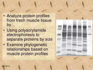
Analyze protein profiles from fresh muscle tissue by… Using polyacrylamide electrophoresis to separate proteins by size Examine phylogenetic relationships based on muscle protein profiles. Traditional classification based upon traits, especially morphological characters.

E N D
Analyze protein profiles from fresh muscle tissue by… • Using polyacrylamide electrophoresis to separate proteins by size • Examine phylogenetic relationships based on muscle protein profiles
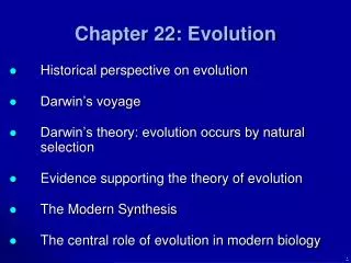
Traditional classification based upon traits, especiallymorphological characters. Traditional Systematics and Taxonomy
DNA RNA Protein Trait Biochemical Similarities • Traits are discrete characteristics. In biology we are concerned with “heritable traits.” • Proteins are generally responsible for traits, most often as enzymes affecting biochemical pathways or as structural or dynamic elements of the cytoskeleton. • DNA codes for proteins that confer traits
Experiment: Compare protein profiles various fresh muscle tissue. • Procedure: • Extract proteins from tissue • Denature proteins • Separate proteins by size using polyacrylamide gel electrophoresis • Stain to visualize protein bands • Analyze and interpret gels
Protein size comparison • Break protein complexes into individual proteins • Denature proteins using detergent and heat • Separate proteins based on size
Acrylamide gel tight matrix Ideal for protein separation Smaller pore size than agarose Proteins much smaller than DNA average amino acid = 110 Da average base pair = 649 Da 1 kilobase of DNA = 650 kDa 1 kilobase of DNA encodes 333 amino acids = 36 kDa Why use polyacrylamide gels to separate proteins?
Protein size • Size measured in kilodaltons (kDa) • Dalton = mass of hydrogen atom = 1.66 x 10 -24 gram • Average amino acid = 110 daltons
Muscle contains proteins of many sizes ProteinkDaFunction titin 3000 center myosin in sarcomere dystrophin 400 anchoring to plasma membrane filamin 270 cross-link filaments into gel myosin heavy chain 210slide filaments spectrin 265 attach filaments to plasma membrane nebulin 107 regulate actin assembly a-actinin 100 bundle filaments gelosin 90 fragment filaments fimbrin 68 bundle filaments actin42form filaments tropomyosin 35 strengthen filaments myosin light chain 27slide filaments troponin (T, I, C) 30, 19, 17 mediate regulation of contraction thymosin 5 sequester actin monomers These are generalized molecular masses and may vary in different species….
Actin and Myosin • Actin: • 5% of total protein • 20% of vertebrate muscle mass • 375 amino acids = 42 kDa • Forms filaments • Myosin: • Tetramer of two heavy subunits (220 kDa) and two light subunits (20 kDa) • Breaks down ATP for muscle contraction
What’s in the sample buffer? • Tris buffer to provide appropriate pH • SDS (sodium dodecyl sulfate)detergent to dissolve proteins and give them a negative charge • Glycerol to make samples sink into wells • Bromphenol Blue dye to visualize samples
Why heat the samples? • Heating the samples denatures protein complexes, allowing the separation of individual proteins by size s-s SDS, heat proteins with SDS - +
Negatively charged proteins move to positive electrode Smaller proteins move faster Proteins separate by size s-s SDS, ß-Me, heat - + proteins with SDS How does an SDS-PAGE gel work?
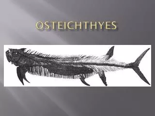
15% SDS-PAGE Lane 1: Marker Lane 2: Shark Lane 3: Salmon Lane 4: Rainbow trout Lane 5: Cod Lane 6: Sturgeon Lane 8: Croaker Lane 9: Weakfish Lane 10: Actin/myosin Gel Analysis
kDa mm Molecular Weight Analysis 203 8.5 135 12.0 86 18.5 41 28.0 33 34.0 19 41.5 8 44.5
Phylogenetic Tree TUNA MACKEREL SALMON TROUT CARP MINNOW SNAPPER PERCH WALLEYE BASS CATFISH COD HAKE POLLOCK SMELT ANCHOVIES HERRINGS SARDINES FLOUNDER SOLE HALIBUT PIKE STURGEON GAR SHARK Agnatha Chondrichthyes Ostheichthyes Amphibia Reptilia Aves Mammalia OYSTER CLAM MUSSEL SCALLOP OCTOPUS SQUID CRAB LOBSTER SHRIMP Mollusk Arthropod Echinoderm Chordate Deuterostome Protostome Metazoa