Download
1 / 12
120 likes | 975 Vues
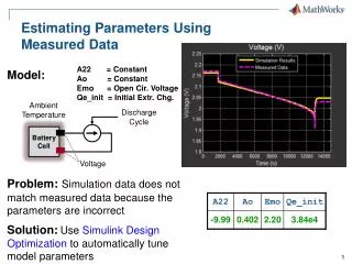
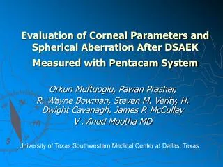
Orkun Muftuoglu , Pawan Prasher , R. Wayne Bowman, Steven M. Verity, H. Dwight Cavanagh, James P. McCulley V . Vinod Mootha MD. Evaluation of Corneal Parameters and Spherical Aberration After DSAEK Measured with Pentacam System.

E N D
Orkun Muftuoglu, PawanPrasher, R. Wayne Bowman, Steven M. Verity, H. Dwight Cavanagh, James P. McCulley V .Vinod Mootha MD Evaluation of Corneal Parameters and Spherical Aberration After DSAEK Measured with Pentacam System University of Texas Southwestern Medical Center at Dallas, Texas
Financial Disclosure • UT Southwestern Medical Center & Aston Ophthalmology Clinic, Dallas, TX. • Drs. Bowman, Verity, and McCulley receive consultant reimbursement from Alcon Inc. • Dr. Cavanagh receives research reimbursements from Ciba Inc and Menion Inc. • None of the authors have financial interest in the subject matter of this poster. • Acknowledgements: Supported in part by an unrestricted research grant from Research to Prevent Blindness, Inc., New York, New York.
Introduction • DSAEK is a technique for selective replacement of dysfunctional endothelium • Rapid recovery of vision • Minimal induced astigmatism and anisometropia • Enhanced resistance to trauma • Less risk of expulsive choroidal hemorrhage • Fewer ocular surface problems • Despite favorable visual outcomes, fewer patients than expected achieve BCVA of 20/20. • Probable limiting factors: subepithelial scarring, interface changes, increased corneal thickness and increased higher order aberrations • Hyperopic shift- Anatomical changes induced to posterior corneal surface???
Purpose • Compare corneal parameters of the eyes that underwent DSAEK with age-matched controls using rotating Scheimpflug imaging system
32 eyes (28 patients) who underwent DSAEK 32 eyes (32 patients) age-matched controls (no previous ocular surgery) Pentacam Study group had Scheimpflug images (Pentacam, Oculus, Germany) taken after 3 months post-operatively Scheimpflug camera and a monochromatic light source The system rotates 180 degrees and takes 25-50 images of the anterior segment in about 2 seconds Calculates a 3-dimensional model of the anterior eye segment from as many as 25,000 true elevation points Paremeters evaluated Mean anterior keratometry (Ka) Mean posterior keratometry (Kp) Mean anterior radius of curvature (Ra) Mean posterior radius of curvature (Rp) True net power (TNP) Central corneal thickness (CCT) Corneal volume (CV) Keratometric Power Deviation (KPD) Mean anterior and posterior astigmatism Equivalent K Readings (EKR) in 2, 4 and 6mm zone HOAs data up to 6th order from anterior and posterior surface in the central 6.0 mm zone Corneal densitometry DSAEK- 16 eyes Phacoemulsification with DSAEK- 14 eyes DSAEK with IOL exchange- 2 eyes 17 clear corneal and 15 scleral tunnel Graft size was 8.0mm or larger in all cases Materials and Methods
Comparison between DSAEK and controls P* = Mann-Whitney U test
Control Anterior Posterior DSAEK VA : 20 / 25
Corneal Densitometry P* = Mann-Whitney U test
Discussion DSAEK surgery- • Creation of a new steeper endothelial surface • Meniscus shaped endothelial graft • Alteration of physiological relationship between anterior and posterior surface • Placido based imaging systems could produce erroneous measurements of corneal power • Measures both anterior and posterior surface • Calculates true net power based on thick lens formula • Incorporates refractive indices at each interface along with anterior and posterior curvature and corneal thickness • Has been shown to be useful in studying changes in posterior curvature and give repeatable measurements of corneal thickness- the parameters most affected in DSAEK Changes induced by DSAEK • Increased corneal thickness and volume • additional stromal tissue • Lower radius of curvature of posterior surface (relative steepening) • Meniscus shaped graft thicker in the periphery • Higher radius of curvature of anterior surface (relative flattening) • Wound related? • Alteration of curvature on resolution of bullous changes?? • Lower true net power in DSAEK corneas
Conclusions • Pentacam measurements show lower true net power in DSAEK as compared to controls likely due to • increased posterior curvature (increased minus lens effect) • Decreased anterior curvature (decreased plus lens effect)?? • Spherical aberration is variable after DSAEK surgery.