Download
1 / 11
110 likes | 343 Vues
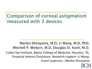
Corneal Spherical Aberration in a Population of Patients Measured by 3 Different Instruments. Mohamed A Guenena, MD Helga P Sandoval, MD, MSCR Kerry D Solomon, MD Magill Research Center for Vision Correction, Storm Eye Institute Medical University of South Carolina, Charleston

E N D
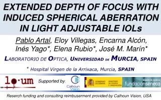
Corneal Spherical Aberration in a Population of Patients Measured by 3 Different Instruments • Mohamed A Guenena, MD • Helga P Sandoval, MD, MSCR • Kerry D Solomon, MD Magill Research Center for Vision Correction, Storm Eye Institute Medical University of South Carolina, Charleston The authors does not have any financial interests in the products mentioned
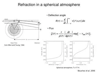
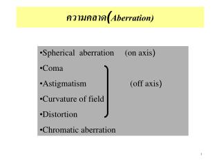
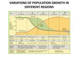
Background • Spherical aberration (SA) is a fourth-order higher-order aberration in which peripheral light rays are refracted more than the central ones • Can be positive or negative depending on where the light rays focuses • Leads to image spread on the retina instead of point focus • Average cornea SA is +.27µ compensated by the lens negative SA in healthy individuals. • After cataract surgery, if a spherical IOL is implanted, it will add more positive SA to the eye
Background • Several aspheric monofocal IOLs have been developed with different SA values to compensate for the corneal SA • Tecnis Z9000 -.27µ • AcrySof SN60WF -.20µ • nanoFLEX Collamer -.019µ • Sofport AO 0 nanoFLEX Sofport AO Tecnis SN60WF
Background • Literature suggests getting the patient closest to 0 SA to achieve the optimum quality of vision • Correct selection of the aspheric IOLs will depend mainly on SA measurements of the cornea
Purpose • To compare the measurements of corneal spherical aberration obtained by the Atlas Corneal Topography (Carl Zeiss, Jena, Germany), Galilei Dual Scheimpflug Analyzer (Ziemer, Port, Switzerland) and the OPD-Scan II (Nidek, Tokyo, Japan). Galilei Dual Scheimpflug Analyzer Atlas Corneal Topography OPD-Scan II
Methods • A pilot study • SA were measured in patients undergoing cataract surgery before dilation. • SA measured in the 6 mm optical zone • No history of ocular surgery or injury • No ocular pathology other than cataract • All measurements were done by single trained observer • Difference between measurements were analyzed
Measurements 8 eyes of 6 patients All females Average age: 73 years ( 63-81)
Difference between Galilei and OPD measurements compared to Atlas Positive sign = overestimate Negative sign = underestimate
Conclusion • This pilot study showed high consistency of measurements of SA among the 3 tested instruments • This finding can help the surgeons choose the proper aspheric IOL regardless of which measuring instrument is used. • More studies are needed to determine the optimal SA surgeons need to target