Physiology of Strength Training
Physiology of Strength Training Part 1 – Muscle Function Skeletal Muscle Anatomy 660 muscles Approximately 45% of body weight 75% water, 20% protein (12% myofibullar and 8% enzymes, etc) 5% inorganic salts Largest organ system End organ for the primary support systems involved in exercise

Physiology of Strength Training
E N D
Presentation Transcript
Physiology of Strength Training Part 1 – Muscle Function
Skeletal Muscle Anatomy • 660 muscles • Approximately 45% of body weight • 75% water, 20% protein (12% myofibullar and 8% enzymes, etc) 5% inorganic salts • Largest organ system • End organ for the primary support systems involved in exercise
Skeletal Muscle Anatomy • Basement membrane – outer most ‘membrane’ • Plasma membrane or sarcolemma • Satellite cells – between membranes • growth, development, adaptation • under stress they are responsible for hypertrophy and hyperplasia • Multi-nuclei • 200-300 nuclei per millimeter • 85-95% within the scaroplasm • 5-15% in the basement membrane • Tremendous potential for gene alterations
Skeletal Muscle Anatomy • Muscle • Fascicle • Fibers
Skeletal Muscle Structure • Epimysium, Endomysium, and Perimysium converge to form tendons and are very elastic
Skeletal Muscle Anatomy • Muscle • Fascicle • Fibers • Myofibrils • Myofilaments • Actin • Myosin
Skeletal Muscle Anatomy • What are the three proteins that make up actin? • Actin, tropomyosin, and troponin • What is the function of actin? • What is the function of tropomyosin? • What is the function of tropoin?
Skeletal Muscle Anatomy • Myosin is also referred to as the thick myofilament • Myosin heads • What does myosin use its heads for?
Skeletal Muscle Anatomy • Myosin • Actin • M-line proteins • Titin/Nebulin
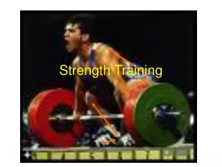
Muscle Strength • Where is the strength of a muscle generated? • Muscle strength comes from the interaction of myosin and active • How is the strength of a muscle generated?
Muscle Contraction:Generating Force • Stimulation • Calcium • Actin/Myosin • Sliding • Energy/ATP
Motor Unit • What is a motor unit? • One motor nerve • All the muscle fibers (cells) that it stimulates • Example. 1:4 • Actual MU range from 1:10s to 1:1000s
Neuromuscular Junction • How does a nerve stimulate a muscle fiber? • Neurotransmitter substance • Acetylcholine (ACh)
Neuromuscular Junction • Acetylcholine (acetate and choline) • Reuptake of choline • Fatigue? • Supplement?
Depolarization and Action Potential • Acetycholine • Sodium gates open • Depolarization • Action Potential • Propagation of Action Potential • Sarcolemma • T-tubules
Muscle Contraction: Calcium • After the T-tubule, where does the action potential go? • What is stored in the sarcoplasmic recticulum? • Where does the calcium go? • FYI: Too much lactic acid may block Ca release.
Muscle Contraction: Actin/Myosin • Ca binds to troponin • Troponin causes tropomyosin to shift or move • This uncovers the active sites on the actin. • Once uncovered, the myosin heads will attach to the active site
Muscle Contraction: Sliding • What does the myosin head do once it attaches? • This is called a power stroke. • What happens to the actin? • The myosin breaks away, reattaches, power stroke… • In this case, what happens to the sarcomere?
Actin and Myosin Interaction Charged myosin head and covered active sites. 1. 2. Uncovered active site; myosin head attaches. 3. Power stroke sliding actin inward; myosin head uncharged. 4 ATP separates and recharges myosin head.
Muscle Contraction: Energy • What is ATP? • What does it provide?
ADP + P Energized ADP + P + energy New ATP
Muscle Relaxation • What is needed in order to have the muscle stop contracting? • Stop the impluse • Re-store the calcium • How? Calcium pump • What powers the calcium pump?
Summary of Muscle Contraction 1. Motor Impulse 2. Neurotransmitter Substance 3. Action potential via Na and K 4. Calcium released exposing active sites 5. ATP split forming high energy myosin
Summary of Muscle Contraction 6. Myosin attaches to actin forming a crossbridge 7. Stored energy released and crossbridge movement (Power Stroke) 8. ATP breaks myosin from actin 9. ATP splits forming high energy myosin
Quick Time Movie • Yellow = Calcium • Green = ATP • Gray = ATPase • A Quick Time Movie of the contraction process can be download at the 5230 Web Page
Muscle Strength • So far… • ...Where and how is the strength of a muscle generated? • Next… • ….What makes a muscle stronger? • Acute increase • Chronic increase