
Tuesday Case of the Day
E N D
Presentation Transcript
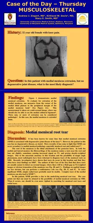
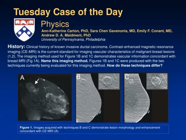
History:Clinical history of known invasive ductal carcinoma. Contrast-enhanced magnetic resonance imaging (CE-MRI) is the current standard for imaging vascular characteristics of malignant breast lesions (1,2). The imaging method used for Figure 1B and 1C demonstrates vascular information concordant with breast MRI (Fig 1A). Name this imaging method. Figures 1B and 1C were produced with the two techniques currently being evaluated for this imaging method. How do these techniques differ? Tuesday Case of the Day Physics Ann-Katherine Carton, PhD, Sara Chen Gavenonis, MD, Emily F. Conant, MD, Andrew D. A. Maidment, PhD University of Pennsylvania, Philadelphia Figure 1. Images acquired with techniques B and C demonstrate lesion morphology and enhancement concordant with CE-MRI (A).
Answers:The new method is contrast-enhanced digital breast tomosynthesis(CE-DBT). In contrast to projection mammography, DBT is an x-ray imaging technique that provides 3D images of the breast (3). DBT is thus able to depict 3D lesion morphology and lesion localization. With the administration of an iodinated vascular contrast agent, it is possible to demonstrate vascular characteristics of breast lesions, as is illustrated in this case. Two techniques for CE-DBT are being studied: temporal subtraction CE-DBT (4) and dual-energy (DE) subtraction CE-DBT (5). Note that the CE-DBT images are single-slice in-plane images of the breast in MLO position.
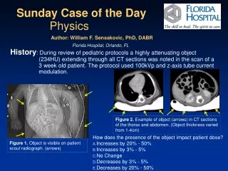
HE HE Iodine contrast Pre contrast Post contrast Time Discussion: Temporal Subtraction CE-DBT K-edge Iodine HE spectrum Breast Figure 2. In temporal subtraction, one precontrast and one or more postcontrast tomosynthesis time points are acquired by using a high-energy (HE) x-ray spectrum. Iodine-enhanced images are produced by subtracting the logarithm of the pre- and postcontrast series (4).
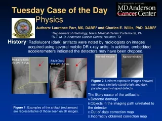
LE HE Iodine contrast LE HE Time Discussion: Dual-Energy Subtraction CE-DBT LE spectrum K-edge Iodine HE spectrum Breast Figure 3. DE CE-DBT requires only postcontrast image series at two energies that closely bracket the K-absorption edge of iodine. Iodine-enhanced images are given by weighted difference between the logarithms of the HE and low-energy (LE) images (5).
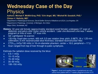
Discussion:Theoretically, in the absence of breast motion, temporal subtraction is the most sensitive method for enhancement detection, since background breast tissue can be completely cancelled, whereas in DE CE-DBT, background tissue cannot be completely cancelled. In clinical application, however, temporal subtraction is very susceptible to breast motion artifacts due to the relatively long time delay between the pre- and postcontrast series (4). With DE CE-DBT, artifacts from breast motion can be significantly reduced because the images of the image series pair at each time point are acquired in rapid succession. Investigation is ongoing into optimizing CE-DBT and comparing temporal CE-DBT with dual-energy CE-DBT techniques. Temporal subtraction DE subtraction Figure 4. Temporal and dual-energy CE-DBT images. Note the more significant motion artifacts in the temporal subtraction CE-DBT image; the clip inside the lesion shows a displacement of approximately 2 mm.
References • Kuhl CK, Schild HH, Morakkabati N. Dynamic bilateral contrast-enhanced MR imaging of the breast: trade-off between spatial and temporal resolution. Radiology 2005;236:789-800. • Schnall MD, Blume J, Bluemke DA, et al. Diagnostic architectural and dynamic features at breast MR imaging: multicenter study. Radiology 2006;238:42-53. • Niklason LT, Christian BT, Niklason LE, et al. Digital tomosynthesis in breast imaging. Radiology 1997;205:399-406. • Chen SC, Carton AK, Albert M, et al. Initial clinical experience with contrast-enhanced digital breast tomosynthesis. Acad Radiol 2007;14:229-238. • Carton AK, Ullberg C, Lindman K, Francke T, Maidment ADA. Optimization of a dual-energy contrast-enhanced technique for a photon counting digital breast tomosynthesis system. 9th International Workshop, IWDM 2008: Tucson, AZ; July 2008.