Download
1 / 22
320 likes | 1.6k Vues
Management of Chondral and Osteochondral defects of Knee joint. Dr Deepak Goyal Consultant Knee Surgeon MS (Orth), DNB (Orth), MNAMS Fellow of Knee Surgery Wellington Knee Hospital, London, UK Semmelweis University, Budapest, Hungary Hinchingbrooke Hospital, Cambridgeshire, UK

E N D
Management of Chondral and Osteochondral defects of Knee joint Dr Deepak Goyal Consultant Knee Surgeon MS (Orth), DNB (Orth), MNAMS Fellow of Knee Surgery Wellington Knee Hospital, London, UK Semmelweis University, Budapest, Hungary Hinchingbrooke Hospital, Cambridgeshire, UK Uzsoki Hospital, Budapest, Hungary
Hunter 1743 “From Hippocrates to present age, it is universally allowed that ulcerated cartilage is a trouble some thing & that once destroyed, is not repaired” Type IV defect
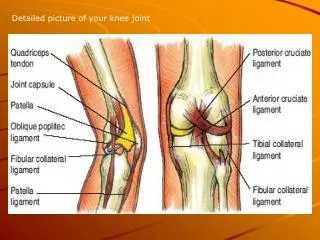
Cartilage • Cartilage is marvelous: tough, elastic, durable. • If normal, it will last lifetime. • But if damaged; even normal activity lead to erosion of joint surface.
Causes of Cartilage injuries • Sports • involving torsional forces, high speed sports. • Trauma • RTA, Bike injuries • Pathological • OCD • Associated with other IA lesions • Neglected • Missed • Ignored.
ICRS Arthroscopic Classification Lesion Thickness • Grade 0: Normal • Grade I: Superficial fissures • Grade II:<1/2 depth
ICRS Arthroscopic Classification • Lesion Thickness • Grade III: >50% depth but not thru subchondral plate. • Grade IV: lesion thru subchondral plate • OCD lesions • AVN lesions
Treatment options • Some light is now seen at the end of ‘centuries old dark tunnel’. • Benign neglect • Debridement • Pridie’s perforations • Abrasio Arthroplasty • Kevin’s Morselized osteochondral mixture • Steadmann’s Microfracture • Periosteal Grafting • Perichondrial Grafting • Osteochondral Allograft • Osteochondral Autograft • Mosaicplasty • ACI • Biomaterials
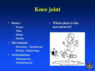
Treatment goals • Permanent restoration of cartilage surface. • Hyaline or Hyaline like regeneration. • Ideal. • If not possible, fibro cartilage regeneration. • Poor biomechanical properties. • Tends to wear off fast. • Doest not allow high demand activities.
Benign Neglect & close monitoring • < 1 cm lesions. • Documentation must. • Close monitoring. • Only short Follow Up available to support. Natural History Type of cartilage: ??Fibrocartilage Life: ??????? Shelbourne et al. American Academy of Orthopedic Surgeons. Annual meeting. 2002.
Debridement and Lavage • Temporary relief because fails to target the primary defect Natural History Type of cartilage: ??Fibrocartilage Life: Temporary Moseley JB et al. N Engl J Med. 2002; 347:81-88.
Pridie’s Perforations • Multiple drilling of subchondral bone. • To stimulate fibrocartilage. • 1st attempt of a systematic approach. • Thermal necrosis of surrounding tissues. Natural History Type of cartilage: Fibrocartilage Life: Not available Pridie KH. A method of resurfacing osteoarthritic knee joints. JBJS. 1959; 41: 618-619.
Johnson’s Abrasion Athroplasty • Abrasion of superficial subchondral bone. • Exposes mesenchyme & intraosseous vessels. • Violates integrity & contour of subchondral bone. • Thermal necrosis Natural History Type of cartilage: Fibrocartilage Life: ?????? Johnson LO. Arthroscopic abrasion arthroplasty, historical and pathological perspective, present status. Arthroscopy. 1986; 2: 54.
Steadman’s Microfracture/ CPM • 81 patients • Age 13-45 years • Average size 2.83 cm2 • Follow Up 11.3 years-compliance 97% • Lysholm Score 58.8>88.9 • Degenerative defect- no contraindication • Size> 4 cm2: less improved Natural History Type of cartilage: Fibrocartilage Life: Upto 10 years in selected conditions Steadman et al. J of Knee surgery: 2004: vol 17.1 Jan: 13-17.
Osteochondral Allograft • Gross 1992 Toronto • 50 patients • 35 years average age • 5 years: 95% Good/ Excellent • 10 years: 71% Good/Excellent • 20 years: 66% Good/Excellent • Bugbee 2000 ICRS • 211 patients • 52 months ave F-U. • 38 years ave age. • 93% Good/ Excellent Natural History Type of cartilage: ?Hyaline or like Life: Upto 20 years in selected conditions Gross et al. JBJS Br. 1997; 79:1008-1013.
Hangody’s Mosaicplasty • Cylindrical Osteochondral grafts • From NWB articular surface. • To WB articular surface. • Indications: • Non degenerative • 1.5 to 4 cm2 • Isolated • Absolute contraindications: • Age > 50 • Size > 4-8 cm2 • Depth > 10 cm. Hangody et al. JBJS-Am. March 2004; 86-A supp 1: 65-72.
Hangody’s Mosaicplasty • 831 cases • 12-13 years of F/U • Good/Excellent results • 92% femoral condyle • 87% in Tibial resurfacing • 79% in patella/ Trochlea • Donor site morbidity: 3% Natural History Type of cartilage: Hyaline or like Life: medium to long term good results Hangody et al. JBJS-Am. March 2004; 86-A supp 1: 65-72.
Autologous chondrocytes implantaion Cost: 11000$ only • Most interesting & controversial treatment. • Two surgical procedures. Natural History Type of cartilage: Hyaline Life: Short to medium good results Peterson L. 2001; 391:S337-S348.
Other techniques • Kevin Stone’s morselized autogenous osteochondral mixture. • Periosteal grafting. • Osteochondral autogenous transfer.
Biomaterials, Hyaluronan based Scaffolds • Osteo-inductive material like BMP. • Chondro-inductive material like Hyaluronan. • Osteocytes and Chondrocytes. Futuristic!!! Type of cartilage: Hyaline Life: ?Whole Life Personal Communication Lazslo Hangody: Budapest. 2004.
Treatment Protocol….Algorithm • Grade 1: Conservative • Grade 2: • Debridement • Extensive controlled rehabilitation • Grade 3 & 4: • < 1cm: Benign neglect or microfracures • 1-2 cm: Microfractures • 1.5 to 4 cm: Mosaicplasty • 3 to 10 cm: ACI, osteochondral allografts
Precautions • Careful selection of patient and treatment option, proper assessment of size and depth of lesion, • can prevent if not all some arthritic knee. • Mal-alignment, meniscal deficiency, cruciate deficiency etc are either the cause of injury or cause of progression of chondral lesions. • Do not forget to correct the biomechanics.
Thank you Dr Deepak Goyal Consultant Knee Surgeon MS (Orth), DNB (Orth), MNAMS Fellow of Knee Surgery • Semmelweis University, Budapest, Hungary • Uzsoki Hospital, Budapest, Hungary • Hinchingbrooke Hospital, Cambridgeshire, UK • Wellington Knee Hospital, London, UK