False positive extra-parathyroid imaging or Ectopic parathyroid gland?
False positive extra-parathyroid imaging or Ectopic parathyroid gland?. Based on a 29 y/o female with primary hyperparathyroidism & extra- parathyroidal uptake in nuclear imaging Presented By: A.H. Ghanooni Fellow of Endocrinology in SBMU July 2017. ANATOMY OF PARATHYROID GLANDS.

False positive extra-parathyroid imaging or Ectopic parathyroid gland?
E N D
Presentation Transcript
False positive extra-parathyroid imaging or Ectopic parathyroid gland? Based on a 29 y/o female with primary hyperparathyroidism & extra-parathyroidal uptake in nuclear imaging Presented By: A.H. Ghanooni Fellow of Endocrinology in SBMU July 2017
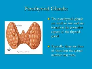
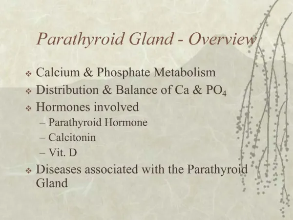
ANATOMY OF PARATHYROID GLANDS • There are normally two pairs of parathyroid glands, the upper and lower pairs, which are located close to the dorsal side of the thyroid gland.
Ectopic parathyroid gland • Approximately 6% of normal individuals have one or more of the glands sited in ectopic positions, • occasionally there may be more than four glands. • Ectopic glands can be found in a wide range of sites but most commonly are in the neck, thymus, carotid sheath and mediastinum. • Glands at any of these sites may become autonomous. • Up to one in five parathyroid glands may be located ectopically, and this is especially true of supernumerary glands. (williams 2017)
determine the incidence of abnormal ectopic parathyroid glands, their specific locations, and the accuracy of preoperative localization using technetium-99m–sestamibi scintigraphy.
Collected from a parathyroid database and a retrospective chart review of consecutive patients operated on for hyperparathyroidism from 1990 to 2005. • Definition : • An ectopic inferior parathyroid gland was defined as a gland in a location other than on or immediately adjacent to the anterior or posteriolateral surface of the inferior pole of the thyroid gland. • An ectopic superior parathyroid gland was defined as a gland in a location other than juxtacricothyroidalposteriorlyor within the capsule of the posterior surface of the superior pole of the thyroid gland.
Results: • Of the 231 patients operated on for hyperparathyroidism, 37(16%) had an abnormal ectopic parathyroid gland. • Ectopic parathyroid glands were superior in 14 (38%) and inferior in 23 (62%) patients.
Results: • Ectopic Superior glands were found in (14 patients) : • tracheoesophageal groove in 6 (43%), • Retroesophageal in 3 (22%), • in the posterosuperiormediastinum in 2 (14%), • intrathyroidal in 1 (7%), • within the carotid • sheath in 1 (7%), • and paraesophageal in 1 (7%) patient. • Ectopic Inferior glands were found in (23 patients): • thymus in 7 (30%), • The anterosuperior mediastinum in 5 (22%), • intrathyroidal in 5 (22%), • the thyrothymic ligament in 4 (17%), • and undescended in a submandibular location in 2 (9%) patients.
LOCALIZATION OF PARATHYROID GLAND • Techniques used include high-resolution ultrasound (US), computerized tomography (CT), magnetic resonance imaging (MRI), selective venous sampling and a variety of nuclear medicine techniques. • The use of high-resolution ultrasonography has reported sensitivities between 43% and 92%. • CT has a reported sensitivity of 35-76% but probably has its major use in examining the mediastinum. • Venous sampling is a relatively invasive technique requiring a high degree of operator dependency and may be useful in persistent or recurrent disease. • MRI has a reported sensitivity of 50-93% and is still undergoing further evaluation. • These techniques therefore have similar ranges of sensitivity of detection, and this is partly determined by the size of the lesion.
RADIONUCLIDE TECHNIQUES • 99mTc-hexakis-2-methoxyisobutyl-isonitrile (99mTc-sestamibi) imaging is now the technique of choice for parathyroid localization. • The difference in retention may be explained by the increased number of mitochondria in the cells of an adenoma and the fact that 99mTc-sestamibi is sequestered in the mitochondria.
FALSE-POSITIVE SCANS • the most common cause of false-positive scans is solid thyroid nodules. • Other false-positive images have been described with sarcoidosis, thyroid carcinoma, lymphoma, and other tumors. • Sestamibi may also be seen to accumulate in brown tumors as well as parathyroid metastases.
A prospective database of 3,187 patients who underwent neck exploration for PHPT was reviewed • to identify patients who had concurrent thyroid resection. • Patients with benign and malignant thyroid disease were comparatively analyzed.
Results: A total of 470 patients underwent both thyroidectomy and parathyroidectomy • Dual-isotope scintigraphyobtained in 374 patients (80%) had a sensitivity of 67% and a positive predictive value of 66% for parathyroid adenoma localization in these patients with thyroid disease. • False-positive scintigraphy occurred in 22% with benign and 45% with malignant thyroid disease (P = 0.002).
Key message • A Tc-MIBI-Hot/I-123-Cold phenotype is very specific for detecting thyroid malignancy. Patients with this imaging phenotype should strongly be considered for preoperative ultrasound-guided biopsy. • Patients found intraoperativelyto have false-positive parathyroid scintigraphy should be evaluated for thyroid cancer.
HPT-JT (HPT- Jaw Tumor Syndrome) • HPT-JT is a syndrome of HPT, jaw tumors, and renal lesions. • Transmission is autosomal dominant. • The most common and sometimes the only feature is HPT. • The HPT typically involves one parathyroid gland at a time, and there is a uniquely high malignant potential in the parathyroid tumor; 15% of patients have parathyroid cancer.
HPT-JT (HPT- Jaw Tumor Syndrome) • The associated jaw tumors (in 25%) are ossifying or cementifyingfibromas. • Unlike the jaw tumors of HPT, they are not osteoclast-rich or influenced by the parathyroid status. • The associated renal lesions (in 5%) are multiple renal cysts, hamartomas, or Wilms tumor. • Uterine tumors are common and can impair fertility.