8.2 Structures and Processes of the Nervous System
UNIT 4. Chapter 8: The Nervous System and Homeostasis. Section 8.2. The nervous system performs the vital role of regulating body processes and structures to maintain homeostasis. The nervous system has two major divisions:. 8.2 Structures and Processes of the Nervous System.

8.2 Structures and Processes of the Nervous System
E N D
Presentation Transcript
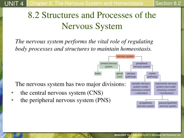
UNIT 4 Chapter 8: The Nervous System and Homeostasis Section 8.2 The nervous system performs the vital role of regulating body processes and structures to maintain homeostasis. The nervous system has two major divisions: 8.2 Structures and Processes of the Nervous System • the central nervous system (CNS) • the peripheral nervous system (PNS)
UNIT 4 Chapter 8: The Nervous System and Homeostasis Section 8.2 The central nervous system (CNS) consists of the brain and spinal cord. The CNS integrates and processes information sent by nerves. The peripheral nervous system includes nerves that carry sensory messages to the CNS and nerves that send messages from the CNS to the muscles and glands (effectors). The peripheral nervous system consists of the autonomic and somatic systems. An Overview of the Nervous System
UNIT 4 Chapter 8: The Nervous System and Homeostasis Section 8.2 The nervous system is composed of two main types of cells: Cells of the Nervous System • neurons: basic structural and functional units of the nervous system. They respond to stimuli, conduct electrochemical signals, and release regulating chemicals. Neurons are organized into tissues called nerves. • glial cells: support neurons by nourishing them, removing wastes, and defending against infection. They also function as structural support cells. Glial cells, shown in green in this micrograph, support neurons (shown in orange).
UNIT 4 Chapter 8: The Nervous System and Homeostasis Section 8.2 In general, neurons share four common features: The Structure of a Neuron • dendrites: short, branching terminals that receive impulses and relay the impulses to the cell body • a cell body: contains the nucleus and is the site of the cell’s metabolic reactions • an axon: conducts impulses away from the cell body and varies in length from 1 mm to over 1 m • branching ends: found on dendrites and axons, they increase the surface area available for receiving and sending information Continued…
UNIT 4 Chapter 8: The Nervous System and Homeostasis Section 8.2 Axons are enclosed in a fatty, insulating layer called the myelin sheath (protects neurons and increases the rate of nerve impulse transmission). They are composed of Schwann cells (a type of glial cell) The Structure of a Neuron
UNIT 4 Chapter 8: The Nervous System and Homeostasis Section 8.2 Structurally, neurons are classified based on the number of processes that extend from the cell body. Classifying Neurons Continued…
UNIT 4 Chapter 8: The Nervous System and Homeostasis Section 8.2 Functionally, neurons are classified as one of three types: Classifying Neurons • sensory neurons: receive input and transmit impulses from sensory receptors to the CNS • interneurons: found in the CNS;link btw. sensory and motor neurons; they process incoming sensory input and relay outgoing motor information • motor neurons: transmit info. from the CNS to effectors (muscles, glands, organs) Continued…
UNIT 4 Chapter 8: The Nervous System and Homeostasis Section 8.2 Classifying Neurons This diagram shows how a sensory neuron, an interneuron, and a motor neuron are arranged in the nervous system. (The breaks indicate that the axons are longer than shown.) Continued…
UNIT 4 Chapter 8: The Nervous System and Homeostasis Section 8.2 -An involuntary reflex action in response to a stimulus. -Allows a rapid response, occurring in about 50 ms. -Brain centres are not activated until after the response. The Reflex Arc
UNIT 4 Chapter 8: The Nervous System and Homeostasis Section 8.2 Neurons use electrical signals to communicate with other neurons, muscles, and glands. The signals, called nerve impulses, involve changes in the amount of electric charge across a cell’s plasma membrane. The Electrical Nature of Nerves
UNIT 4 Chapter 8: The Nervous System and Homeostasis Section 8.2 In a resting neuron, the cytoplasmic side of the membrane (inside the cell) is negative, relative to the extracellular side (outside the cell). This charge separation across the membrane is a form of potential energy called membrane potential. (-70mV) Resting Membrane Potential Continued…
UNIT 4 Chapter 8: The Nervous System and Homeostasis Section 8.2 The process of generating a resting membrane potential of -70 mV is called polarization. Resting Membrane Potential cont’d Continued…
UNIT 4 Chapter 8: The Nervous System and Homeostasis Section 8.2 Resting Membrane Potential cont’d…
UNIT 4 Chapter 8: The Nervous System and Homeostasis Section 8.2 The sodium-potassium pump is the most important factor that contributes to the resting membrane potential. This system uses ATP to transport 3 Na ions (Na+) out and 2 potassium ions (K+) into the cell. The overall result of this process is a constant membrane potential of -70 mV. Sodium-Potassium Pump
UNIT 4 Chapter 8: The Nervous System and Homeostasis Section 8.2 Recall that a neuron is polarized due to the charge difference across the membrane. Depolarization occurs when the cell becomes less polarized (the membrane potential is reduced to less than the resting membrane potential of -70 mV). During depolarization, the inside of the cell becomes less negative relative to the outside of the cells. An action potential causes depolarization to occur. Action Potential Continued…
UNIT 4 Chapter 8: The Nervous System and Homeostasis Section 8.2 An action potential is the movement of an electrical impulse along the plasma membrane of an axon. It is an “all-or-none” response. If a stimulus causes the axon to depolarize to a certain level (the threshold potential), an action potential occurs. Threshold potentials are usually close to -50 mV. Note: The strength of an action potential does not change based on the strength of the stimulus. Action Potential Continued…
UNIT 4 Chapter 8: The Nervous System and Homeostasis Section 8.2 Action Potential The graph shows the changes that occur to membrane potential as an action potential travels down an axon.
UNIT 4 Chapter 8: The Nervous System and Homeostasis Section 8.2 An action potential is triggered when the threshold potential is reached. Action Potential: Step 1
UNIT 4 Chapter 8: The Nervous System and Homeostasis Section 8.2 Voltage-gated sodium (Na+) channels open when the threshold potential is reached. Sodium ions move down their concentration gradient and rush into the axon, causing depolarization of the membrane. The membrane potential difference is now +40 mV. Action Potential: Step 2
UNIT 4 Chapter 8: The Nervous System and Homeostasis Section 8.2 Voltage-gated sodium channels close due to change in membrane potential. Voltage-gated potassium (K+) channels open. Potassium ions move down their concentration gradient and exit the axon, causing the membrane to be hyperpolarized to -90 mV. Action Potential: Step 3
UNIT 4 Chapter 8: The Nervous System and Homeostasis Section 8.2 Voltage-gated potassium channels close. The sodium-potassium pump and naturally occurring diffusion restore the resting membrane potential of -70 mV. The membrane is now repolarized. Action Potential: Step 4
UNIT 4 Chapter 8: The Nervous System and Homeostasis Section 8.2 After an action potential occurs, the membrane cannot be stimulated to undergo another action potential. This brief period of time (usually a few milliseconds) is called the refractory periodof the membrane. The events that occur in an action potential continue down the length of the axon until it reaches the end, where it initiates a response at the next cell. Action Potential
UNIT 4 Chapter 8: The Nervous System and Homeostasis Section 8.2 The junction between two neurons, or between a neuron and an effector, is called a synapse. Neurons are not directly connected. They have a small gap between them called the synaptic cleft. A nerve impulse, however, cannot jump from one neuron to another across the cleft. How does the nerve impulse proceed from the presynaptic neuron (sends out the info.) to the postsynaptic neuron ( receives the info)? Chemical messengers, neurotransmitterscarry the nerve impulse across the synapse from one neuron to another. Signal Transmission Across a Synapse
UNIT 4 Chapter 8: The Nervous System and Homeostasis Section 8.2 The nerve impulse travels to the synaptic terminal. Signal Transmission Across a Synapse: Step 1
UNIT 4 Chapter 8: The Nervous System and Homeostasis Section 8.2 Synaptic vesicles containing neurotransmitters move toward and fuse with the presynaptic membrane. Signal Transmission Across a Synapse: Step 2
UNIT 4 Chapter 8: The Nervous System and Homeostasis Section 8.2 Synaptic vesicles release neurotransmitters into the synaptic cleft by exocytosis. Neurotransmitters diffuse across the synapse to reach the postsynaptic neuron or the cell membrane of an effector. Signal Transmission Across a Synapse: Step 3
UNIT 4 Chapter 8: The Nervous System and Homeostasis Section 8.2 Neurotransmitters bind to specific receptor proteins on the postsynaptic membrane. The receptor proteins trigger ion channels to open. Depolarization of the postsynaptic membrane occurs, and an action potential is initiated if the threshold potential is reached. Signal Transmission Across a Synapse: Step 4
UNIT 4 Chapter 8: The Nervous System and Homeostasis Section 8.2 Neurotransmitters have either excitatory or inhibitory effects on the postsynaptic membrane. Excitatory molecules, like acetylcholine, cause action potentials by opening sodium channels. Inhibitory molecules cause potassium channels to open, causing hyperpolarization. Neurotransmitters