Overview
In just one hour.... pulmonary thromboembolic diseasepulmonary hypertensionno time to discussalveolar capillary membrane and pulmonary edemapulmonary arteriovenous malformationshemoptysis - small group case. Pulmonary thromboembolism-a huge problem. most common pulmonary disorder among hospitalized patientspulmonary emboli effect at least 650,000 people each year in the USpulmonary emboli account for 100,000 - 200,000 deaths each year.

Overview
E N D
Presentation Transcript
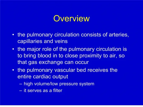
1. Overview the pulmonary circulation consists of arteries, capillaries and veins
the major role of the pulmonary circulation is to bring blood in to close proximity to air, so that gas exchange can occur
the pulmonary vascular bed receives the entire cardiac output
high volume/low pressure system
it serves as a filter
4. Pulmonary thromboembolism-a huge problem most common pulmonary disorder among hospitalized patients
pulmonary emboli effect at least 650,000 people each year in the US
pulmonary emboli account for 100,000 - 200,000 deaths each year
5. Sources of pulmonary emboli thrombi that form in the venous circulation
propagate (grow)
dislodge and travel through the central veins to the pulmonary arteries
femoral, iliac and pelvic veins are the major sources for clinically important pulmonary emboli
subclavian veins, dilated right ventricle are less common sources
6. Factors leading to thrombus formation stasis
abnormal coagulation
enhanced coagulation
impaired fibrinolysis
tissue injury
release of coagulant factors
endothelial cell dysfunction
7. Conditions predisposing to venous thromboembolism Surgery - orthopedic procedures (especially hip repair), urologic procedures
Trauma
Malignancy
Pregnancy
Oral contraceptives
Hormone replacement therapy
Myocardial infarction
Congestive heart failure
Extended bed rest/long periods of travel
Nephrotic syndrome
Antiphospholipid antibody syndrome
Hyperhomocysteinemia Genetic abnormalities in coagulation/fibrinolysis
Deficiency of antithrombin III
Deficiency of protein C
Deficiency of protein S
Factor V Leiden mutation (activated protein C resistance)
Other genetic processes
Sickle cell disease
8. Risk factors in patients with venous thromboembolic disease predisposing factor(s) can eventually be identified in the majority (perhaps > 80%) of patients with venous thromboembolism
prior thromboembolism confers significant risk for subsequent events
more than one risk factor ? further increased risk
over half the episodes in individuals with hereditary coagulation abnormalities occur in the setting of additional stress
9. Circulatory effects of PE clots that lodge in the pulmonary arteries occlude portions of the pulmonary capillary bed
the pulmonary capillary bed has enormous reserve
with exercise flow can increase 5-fold without increasing pulmonary artery pressure
surgical removal of 50% of the pulmonary circulation without increasing PA pressure
loss of vascular bed to emboli causes far greater circulatory disruption than expected from the fraction of circulation blocked
10. Circulatory effects of PE emboli lead to pulmonary vasoconstriction and disproportionate increase in pulmonary vascular resistance
soluble mediators released from platelets
autonomic reflexes
large emboli ? ?? pulmonary vascular resistance ? right ventricular failure ? inadequate left ventricular filling ? hypotension and shock
smaller, peripheral emboli may result in pulmonary infarction
11. Ventilatory effects of PE
12. Hypoxemia in PE most patients present with moderate hypoxemia- why?
increased blood flow through regions of physiologic shunt
V/Q mismatch due to perfusion of constricted lung units
loss of pulmonary surfactant in areas of pulmonary infarction
reduced mixed venous O2 content due to reduced cardiac output
13. Clinical presentation of PE With massive emboli (>50% of the vascular bed obstructed) patients may present with sudden death or circulatory collapse
14. Physical exam signs in PE
15. PFT�s in pulmonary emboli rarely performed in acute PE
spirometry and lung volumes generally show little or no abnormality
may see mild bronchoconstriction or mild restriction
pleuritic pain and splinting can limit study
DLCO usually reduced due to obstruction of vascular bed
16. Arterial blood gases in PE moderate hypoxemia with widened A-a O2 gradient
not universal
increased ventilation leading to ? PCO2 and ? pH
exception in patients who can not increase minute ventilation
17. EKG findings in PE Sinus tachycardia
Atrial arrhythmias
Right bundle branch block - right ventricular overload
Classic SIQ3T3 pattern
18. Chest radiograph in PE Normal
Low lung volumes
Small pleural effusion
Atelectasis
Decreased vascular markings in an area of lung (Westermark�s sign)
Wedge-shaped infiltrate extending to the pleural surface (Hampton�s hump)
19. Diagnostic studies for PE PE is very common and potentially life threatening
the presenting symptoms, signs and routine studies are nonspecific
the clinician needs a high index of suspicion
ideal diagnostic study for PE
image pulmonary vascular
have high sensitivity and specificity
low morbidity
inexpensive
20. Pulmonary angiography advantages
the �gold standard�; images pulmonary artery very effectively
allows measurement of pulmonary artery pressure
disadvantages
invasive
administration of intravenous radiocontrast
expensive
operator time/availability
21. Radionuclide lung scan perfusion scanning: the patient is injected in a vein with radiolabeled macroaggregated albumin (technetium 99)
labeled aggregates are trapped in pulmonary arterioles; retained thoracic radioactivity is imaged with a camera
sensitive for decreased flow to areas of the pulmonary vascular bed - not specific
22. Radionuclide lung scan ventilation scanning: the patient inhales a gas mixture containing a different radiotracer (xenon 133)
areas of vascular obstruction should have loss of perfusion but preservation of ventilation
processes such as pneumonia, COPD, obstructed large airway present as matched ventilation and perfusion defects
23. Radionuclide lung scan - interpretation V/Q scans provide estimates of the probability that a patient has a PE
the level of clinical suspicion for PE must be taken into account
normal perfusion scan excludes the diagnosis of PE
high probability scan with high clinical suspicion gives >95% likelihood of PE
multiple segmental unmatched perfusion defects
intermediate or indeterminant scans are much less helpful
24. V/Q scans and the likelihood of PE
26. Normal Ventilation Scan
27. Normal Perfusion Scan
28. Abnormal Perfusion Scan with Multiple Defects Secondary to Pulmonary Emboli
29. Other noninvasive tests Because most PE originate as lower extremity DVT, tests for lower extremity DVT are useful if positive - may support therapy without invasive studies
Doppler ultrasound
Impedance plethysmography
tests for enhanced clot degradation - if these prove to be sufficiently sensitive, a negative test may greatly decrease the likelihood of thromboembolism
D-dimers
30. Diagnostic algorithm History and exam
laboratory studies (ABG), CXR and EKG
V/Q scan
if intermediate, consider noninvasive studies of lower extremities
if significant clinical suspicion and neither V/Q nor noninvasive studies are diagnostic, proceed to pulmonary angiogram
31. Helical CT scan for PE high speed CT scanners
bolus radiocontrast injection
excellent definition of main, lobar and even segmental pulmonary arteries
may provide bonus information about the lungs and mediastinal structures
34. Role of helical CT in current practice Likely to be an important addition - a major modality in current practice at U of M
positive and negative predictive values in actual use are still being determined
national multicenter trial now underway
35. Treatment of pulmonary embolism prevention
prophylactic anticoagulants, ambulation, pneumatic compression stockings in patients at high risk of DVT
prompt treatment: heparin, heparin, heparin
supportive therapy with oxygen, fluids
heparin prevents clot formation; does not lyse clot
mortality of untreated pulmonary embolism >30%
mortality after initiation of heparin <2%
36. Low molecular weight heparin problems with unfractionated heparin
short half-life: continuous infusion required
variability requiring frequent laboratory studies
low molecular weight heparin-(enoxaparin, dalteparin)
longer half-life: twice daily subcutaneous injections
standard dosing; no requirement for frequent lab monitoring
stable patients without great physiologic compromise can be managed at home
37. Long term care of the patient with DVT/PE heparin for 5-7 days
longer term anticoagulation with an oral drug
warfarin (coumadin)
duration of anticoagulation
6 months if reversible cause
up to life-long if persistence of predisposing high level risk factors or if recurrent DVT/PE
recent data suggests benefit of life-long low dose anticoagulation
usual outcome is restoration of normal V/Q and PVR
38. Additional therapeutic modalities for PE thrombolytic agents - �Drano�
streptokinase
urokinase
tissue plasminogen activator
embolectomy - �plumber�s helper�
39. Inferior vena cava filter block passage of further clots from legs to lung
excellent for patients failing or not tolerating anticoagulation (bleeding problems)
increased risk of subsequent deep vein thrombosis
work best in conjunction with anticoagulation
41. Pulmonary hypertension the normal pulmonary circulation is a low resistance circuit
enormous capacity to recruit unperfused vessels
distensible vessels and ready recruitment allow greatly increased flows with exercise without increasing resistance across the pulmonary vascular bed
42. Resistance across a vascular bed Resistance is
directly related to the pressure drop across the vascular bed
inversely related to the flow through that bed
43. Pulmonary hemodynamics with exercise
44. Pulmonary hypertension
45. Increased pulmonary vascular resistance vasoconstriction
chronic hypoxemia: high altitude or chronic respiratory illnesses
COPD
pulmonary fibrosis
obstructive sleep apnea
vascular obstruction
recurrent/unresolved pulmonary emboli
schistosomiasis
46. Increased pulmonary vascular resistance increased pulmonary blood flow (left-to-right shunts)
with chronically increased flow there is remodeling of the arteriolar wall
destruction of pulmonary vascular bed
emphysema
pulmonary fibrosis
pulmonary vasculitis
47. Primary pulmonary hypertension idiopathic disorder most commonly seen in women 20-45 years old
pathological changes in the pulmonary arteriolar wall
medial hypertrophy
intimal proliferation and fibrosis
similar pattern in pulmonary hypertension due to specific causes
fenfluamine/dexfenfluramine use for weight loss
chronic cocaine use
HIV
liver disease with portal hypertension
48. Familial primary pulmonary hypertension
49. Lessons from familial PPH At least 10% of PPH is familial
Bone morphogenetic protein receptor II
mutation in BMPR2 associated with familial PPH
member of TGF-� receptor family
a significant portion of patients with sporadic PPH now are found to have mutations in BMPR2 as well
50. Symptoms of pulmonary hypertension symptoms of an underlying disease process may predominate
dyspnea on exertion
fatigue
syncope
chest pain
peripheral edema
51. Signs of pulmonary hypertension increased pulmonic component of the second heart sound (P2)
right ventricular lift/heave
elevated jugular venous pulsations
distended liver
peripheral edema
right ventricular S3 gallop
what about lung sounds?
52. Studies in patients with pulmonary hypertension ECG: right ventricular hypertrophy
CXR: dilated main pulmonary arteries/pruning of peripheral vascular markings
ABG: hypoxemia with exertion
PFT�s: findings c/w underlying disease; decreased DLCO
Echocardiogram
Right heart catheterization
53. Diagnostic approach to pulmonary hypertension careful history and exam, serologies for collagen vascular diseases
ECG and echocardiogram
aggressively identify treatable causes of secondary pulmonary hypertension
hypoxemia (at rest or at night, with sleep apnea)
chronic thromboembolic disease
right heart catheterization ? pulmonary angiography
54. Treatment of pulmonary hypertension treat underlying disease
oxygen - minimize hypoxic vasoconstriction
long term anticoagulation (even in PPH)
vasodilators
Calcium channel blockers
Prostacyclin
Endothelin receptor blockers (Bosentan)
transplantation