Download
1 / 58
710 likes | 2.77k Vues
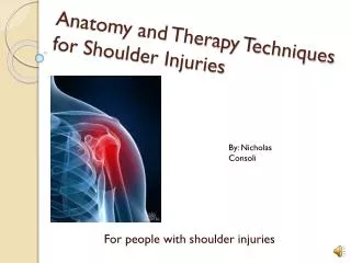
X-Ray Rounds: (Plain) Radiographic Evaluation of the Shoulder. Garry W. K. Ho, M.D. Sports Medicine Fellow - VCU / Fairfax Family Practice December 2006. Anatomy. 3 Bones Humerus Scapula Clavicle 3 Joints Glenohumeral Acromioclavicular Sternoclavicular 1 “Articulation”

E N D
X-Ray Rounds: (Plain) Radiographic Evaluation of the Shoulder Garry W. K. Ho, M.D. Sports Medicine Fellow - VCU / Fairfax Family Practice December 2006
Anatomy • 3 Bones • Humerus • Scapula • Clavicle • 3 Joints • Glenohumeral • Acromioclavicular • Sternoclavicular • 1 “Articulation” • Scapulothoracic
Anatomy • Humerus • Head * • Anatomic neck • Surgical neck • Greater tubercle* • Lesser tubercle* • Intertubercular groove • Deltoid tuberosity • Shaft *
Anatomy • Scapula • Body • Ventral (Costal) surface • Dorsal surface • Borders • Superior • Lateral (Axillary) • Medial (Vertebral) • Angles • Superior • Inferior • Lateral (Head)
Anatomy • Scapula • Glenoid • Acromion • Coracoid • Subscapular fossa • Scapular spine • Supraspinatus fossa • Infraspinatus fossa • Great scapular notch • Suprascapular notch
Anatomy • Scapular “Y” (Lateral)
Anatomy • Clavicle • First bone to start ossification; last to finish • The only bony strut b/w UE and axial skeleton • Flat outer (lateral, acromial) third • Traps, Delt, AC / CC ligaments • Tubular medial (inner, sternal) third • Strongest in axial load • Middle third • Most vulnerable to Fx
Anatomy • Glenohumeral joint • Ball and socket • Purpose: placement of primary prehensile limb • Very mobile; majority (0-120°) of shoulder movement (0-180°) • Price: instability • 45% of all dislocations • Joint stability depends on multiple factors
Anatomy • Glenohumeral joint • Passive stability • Joint conformity • Vacuum effect of jt vol • Synovial fluid adhesion and cohesion • Scapular inclination • Glenoid labrum (50%) • Coracoid ligaments • CCL, CAL • Joint capsule • Glenohumeral ligaments • SGHL, MGHL, IGHLC • Bony restraints • Glenoid fossa, Acromion, Coracoid • Coracohumeral ligament
Anatomy • Glenohumeral joint • Active stability • Biceps (long head) • Rotator cuff • Pectoralis muscles, trapezius, serratus anterior, rhomboids, levator scapulae, etc. (NOT deltoid)
Anatomy • Acromioclavicular joint • Diarthrodial joint • Thin capsule • AC ligaments • Anterior, posterior, superior, inferior • Coracoacromial ligament • Coracoclavicular ligaments • Trapeziod ligament • Conoid ligament
Anatomy • Sternoclavicular joint • Diarthrodial joint • Joint capsule • Articular disk • Intraarticular disk ligament • Sternoclavicular ligaments • Anterior, posterior • Interclavicular ligament
Anatomy • Coordinated shoulder motion • Glenohumeral motion • Acromioclavicular motion • Sternoclavicular motion • Scapulothoracic motion Scapular-humeral rhythm
AP View of the Shoulder • “Transthoracic,” or “Routine” AP View • AP relative to thorax • Suboptimal view of Glenohumeral joint • Good view of AC joint • “Scapular,” “Grashey,” or “Glenohumeral” AP View • Better visualize bony relationships incl GH joint • Suboptimal view of AC joint • Both have been called “True” AP Views
AP View of the Shoulder • “Routine” AP View • Clavicle • Scapula • Acromion & scapular spine • Coracoid • Borders & angles • AC & SC joints • Glenoid • Both ant & post lips • May obscure HH • Humerus • Head & necks • Gr & Lsr tuberosities
AP View of the Shoulder • “Glenohumeral,” “Grashey,” or “Scapular” AP View • Same structures • AC joint not visualized as well • Better visualize the glenoid & humeral head (especially with ER view)
AP View of the Shoulder • AP View in External Rotation • Greater tuberosity & soft tissues profiled and better visualized • Best w/ Scapular AP • AP View in Internal Rotation • May demonstrate Hill-Sachs lesions • GH instability • Best w/ Routine AP
Which AP view should I get? • Routine AP with humeral head in internal rotation (IR) • Scapular / Glenohumeral AP with humeral head in external rotation (ER) Harding WG, Nowicki KD. Plane talk about shoulder radiographs. Phys Sportsmed 1998; 26(2)
Transthoracic Lateral View of the Shoulder • Not usually done • Not as useful • Many obscuring over- and underlying structures
Axillary Lateral View of the Shoulder • Good view of anterior-posterior relationship of GH joint • Coracoid • Acromion • Humerus • Glenoid • GH joint
Axillary Lateral View of the Shoulder • Alternate Axillary views 45° Velpeau View – magnified axillary view
Scapular “Y” Lateral View of the Shoulder • Relationship b/w humeral head and glenoid • Acromion • Coracoid • Scapular body • Scapular spine
Scapular “Y” Lateral View of the Shoulder • Scapular outlet view • A variation of scapular Y view • Same projection, but with beam tilted 5-10° caudad • Shoulder impingement: to evaluate the subacromial space and the supraspinatus outlet
Indications • American College of Radiology (ACR) Appropriateness Criteria for Musculoskeletal Imaging in Shoulder Trauma • Developed in 1995, revised in 2005 • AP with IR & ER, and lateral (axillary or scapular Y) views recommended for: • R/O fracture or dislocation • Subacute (~3 months) shoulder pain suspicious for: • Bursitis / tendonitis • RTC tear or impingement (as initial study)
Indications • Stevenson and Trojian: JFP in July 2002 • No definitive studies on the needs of shoulder radiographs have been done • Recommended obtaining plain films for: • Decreased ROM (especially: abduction < 90°) • Severe pain • History of trauma • Glenohumeral AP, outlet & axillary lateral views • Add AP with IR & ER in cases of trauma • AC joint views for suspected AC joint disease • Neck, chest, abdominal imaging for suspected referred pain Stevenson JH, Trojian T. Applied evidence: evaluation of shoulder pain. J Fam Pract 2002; 51(7):605-611.
Indications • Other indications • Suspicion of instability • Weakness of shoulder motions • The patient cannot communicate (altered mental status, alcohol intoxication, or other) • Persistent pain and decreased ROM • Anytime your history and physical don’t give you enough information
Normal routine AP in IR Normal routine AP in ER Normal axillary view
Routine AP and axillary views Neer classification 3-part proximal humerus fracture involving: - Surgical neck - Lsr tuberosity Tx: surgical eval
Proximal Humerus Fractures:Neer Classification • 2-part fractures • May be Tx’d conservatively if: • Displaced < 1 cm • Angulation < 45 ° • No dislocations • Good reduction • No intraarticular involvement • Anatomic neck intact • Otherwise: surgical evaluation • All else: surgical evaluation
Routine AP in ER, axillary, & scapular Y views Anterior-inferior dislocation No fracture Tx: Conservative
Routine AP in ER, axillary, & scapular Y views Bulb sign, rim sign, loss of parallelism Posterior dislocation; No fracture Tx: Conservative
Post-reduction AP film Routine AP view Inferior GH dislocation (Luxatio erecta) - Rare Tx: may attempt CR
Routine AP in IR and axillary lateral views No dislocation + concave osseous impression in postero-lateral aspect of humeral head What is this lesion called? Hill-Sachs lesion Tx: conservative vs. operative
Type I: conservative tx Type II: conservative tx Type III: conservative tx for most; may consider surgery for active heavy laborers, frequent overhead activity, athletes 20-25 y/o Type IV-VI: surgery Type III AC separation Tx: conservative mostly
Clavicle Fractures • Mostly conservative treatment • Consider surgery for: • Group II Fx’s (esp if medial to CCL) • Open fractures • Neurovascular compromise • Severe associated injuries • E.g. flail chest, mult rib fx’s, scapulothoracic dissociation • Nonunion / malunion
Scapular Fractures • Mostly conservative treatment • Surgical indications: • Controversial • Displaced intraarticular fx’s involving > 25% articular surface • Scapular neck Fx’s with • > 1 cm medial displaced • Angulation > 40 ° • Concomitant fx’s of clavicles, coracoid, acromion, scapular spine • Fracture-dislocations
Routine AP and Axillary Lateral Views Advanced L shoulder osteoarthritis Tx: Symptomatic relief PT / Rehab exercises Injections Consider surgical eval
Scapular Y views A: normal B: Fracture / anterior dislocation C: Posterior dislocation
Routine AP, “True” AP, and Axillary lateral views Split fracture of humeral head with dislocated GH joint Tx: Surgerize!
34 y/o M with shoulder pn and “it feel like it wants to go out of socket” Glenohumeral AP & Scapular Y Lateral views of R shoulder Multiple radiodense loose bodies (largest infra coracoid & infra glenoid) Dx: Loose Bodies Tx: Surgical consult
Glenohumeral AP view of shoulder and humerus Radiolucent lesions spanning proximal third of L humerus Enchondromas Tx: Surgical consult (Biopsy)
Routine AP of R shoulder Group 2, type 2 R clavicle fracture Tx: Surgical repair
Glenohumeral AP, axillary lateral, and scapular Y views Normal findings Tx: as per clinical setting
Routine AP view Scapular body fracture Tx: mostly conservative
Routine AP view Proximal humeral shaft fracture Glenohumeral dislocation Tx: Orthopaedic consult