Download

1 / 10
100 likes | 128 Vues
Looking for Neck Pain Relief? Dr. Amod Manocha specialise in neck pain treatment in delhi and gurgaon.

E N D
Dr. AmodManocha is a Senior Consultant and Head of Pain Management Services at Max Multispecialty Hospital, Saket. He is trained as a Pain Management Specialist and an Anaesthetist in the UK. He has over 13 years of work experience in the UK including working as a Chronic Pain Consultant in many UK hospitals. Phone : +91-987-187-4003 Email: info@removemypain.com Website: http://www.removemypain.com
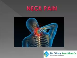
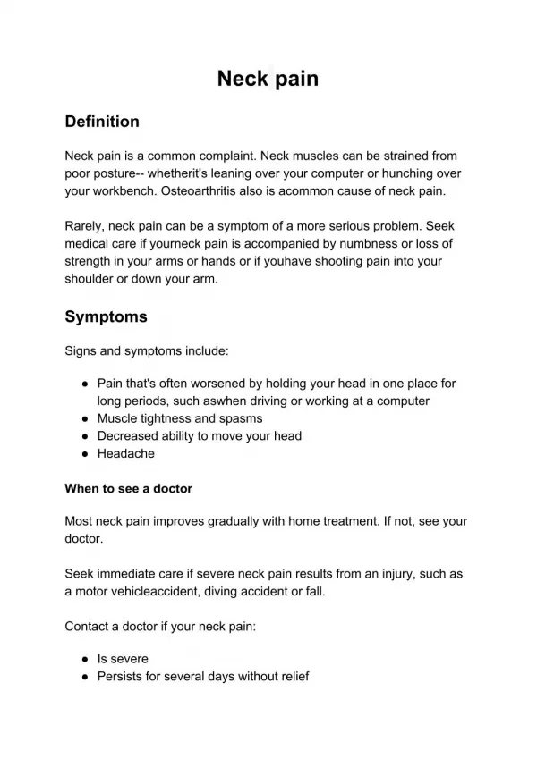
Neck Pain Treatment in Delhi Neck Pain is a pain posteriorly anywhere between the skull base and thoracic spine. It is the largest cause of musculoskeletal disability after low back pain. Approximately two-thirds of the population will suffer from neck pain at some time in their life with high prevalence in middle ages. Fortunately for most people the acute pain resolves within days or weeks although in some it may reoccur or become chronic. Neck pain may be a result of... • Local pathology • Whiplash (flexion-extension) injuries/ trauma • Be a part of a more widespread systemic problem such as ankylosingspondylitis, rheumatoid arthritis, fibromyalgia etc. • Be a result of referred pain from neighboring areas for example the shoulder joint
Facet Joint Injections Spine has many vertebrae and small joints called facet joints link them to each other. The main function of these joints is to provide stability while allowing some degree of movement. These joint commonly become painful and stiff as a result of wear and tear, inflammation or injury. The resulting pain is generally described as a dull ache, heaviness that can radiate towards head, shoulder and scapula. Investigations such as x-rays and MRI may or may not show joint changes. MRI findings alone cannot be relied on to make the diagnosis, as every arthritic joint is not painful. A more reliable test to determine if these joints are responsible for your pain is accurately placed neck injections and if the pain reduced significantly then these joints are the likely source of pain.
Third Occipital Nerve Block& Radiofrequency The third occipital nerve originates from the cervical spine and supplies sensation to a joint in the neck (C2-3 zygapophyseal joint) and a small area at the back of the head. This nerve or the joint it supplies can be a source of headaches localised to the back of head on one side. Sometimes the pain can spread towards the top of the head. This occurs more commonly after whiplash injury. A diagnostic block involving injection of local anaesthetic close to the nerve can help determine if this nerve is the source of your headaches. This is performed under x-ray guidance. If the diagnostic test is positive then radiofrequency ablation of the nerve can provide long lasting relief.
Epidural Epidural space is present in the spine around the sac containing the spinal cord and the nerves. It extends from the back of head to the bottom of spine. Epidural injection involves placing a needle in this space under x-ray guidance. A dye (contrast agent) is used to confirm needle placement before a mixture of local anaesthetic and steroid is given. The level at which the injection is performed will depend on the pathology site and the pain distribution. Depending on the level at which the epidural injection is performed, it may be termed as Cervical epidural – Indicated for neck and arm pain. It involves performing an injection at the base of neck under x-ray guidance Thoracic epidural - Indications for this injection include mid back, chest or abdominal pain. It is performed at mid back level between the shoulder blades, under x-ray guidance. Lumbar epidural is performed for back, groin or leg pain. It involves a x-ray guided injection in the lower back at the waist level Caudal epidural is performed for back and leg pain. It involves a x-ray or ultrasound-guided injection close to the lower end of the spine near the tailbone
Stellate Ganglion Block & Radiofrequency Stellate ganglion is a collection of special type of nerves called sympathetic nerves lying at the base of the neck. These nerves control blood flow, sweating, temperature sensation and are sometimes involved in transmitting pain. Stellate ganglion block interrupts the flow of signals in these nerves and as a consequence reduces pain and increases blood flow to the arm. These injections are used in conditions when sympathetic nerves are involved in transmitting pain such ascomplex regional pain syndrome, post herpetic neuralgia (PHN) and in conditions with reduced arm blood supply such raynaud's syndrome, frostbite. This injection is performed for diagnostic or therapeutic reasons using ultrasound or x-ray. The effects from the injection can help streamline further treatment plan and provide an opportunity to interrupt the pain cycle, engage in physical therapy. Pulsed radiofrequency treatment can help in prolonging the benefits if the effects of block are short lasting.
Nerve Blocks • Occipital nerves originate from the neck and travel to the back of the head and scalp. On route the nerves are liable to compression at points where they cross different muscle planes. Compression or trauma to these nerves gives rise to occipital neuralgia which presents as a burning, shooting, aching pain at the back of head. It may also result from degenerative changes or nerve compression at the level of cervical spine. • Occipital Nerve block is performed using ultrasound guidance. It can be performed at the back of head however sometimes a deeper injection is required in cases of proximal entrapment. A mixture of local anaesthetic and steroid is injected and the spread of the medications can be directly visualised when using ultrasound guidance. Pulsed radiofrequency can help to prolong the analgesic effect.
Atlantoaxial Joint Injection These are joints between the first and the second vertebrae of the spine. These joints can be affected by degenerative and osteoarthritis changes giving rise to headaches or pain localised to the base of skull / top of the neck along with limited neck rotation. Injection of this joint can be performed under x ray guidance. These have to be performed carefully due to proximity of important structures such as spinal cord and blood vessels supplying the brain (vertebral artery). The injection involves checking for needle position using a dye (contrast) followed by injection of a mixture of local anaesthetic and steroid.
Trigger point injections Skeletal Muscles form a substantial proportion of human body and their ability to contract and relax helps in producing body movements. When muscles fail to relax, they form knots or tight bands known as trigger points. These can be a result of inflammation, trauma and injury of the muscle or the neighbouring structures. Poor posture and repetitive strain are other predisposing factors. They are more commonly observed in trapezius, neck and lower back muscles. Pressure over a trigger point produces local soreness and may refer pain to other body parts. Trigger points can limit the range of movement; affect posture predisposing other areas to unaccustomed strain. Trigger point injections are performed in an outpatient/ day-care setting and involve injection of a mixture of local anaesthetic and steroid. I prefer to perform these injections under ultrasound guidance as this helps in improving the accuracy and reduces the chances of complications. Post injection physiotherapy is essential to prevent recurrence and maximise the benefits.
Common Musculoskeletal Conditions Tennis/Golfer’s Elbow These are common problems involving staining/overuse of forearm tendons which attach to the bony prominences on the upper arm bone close to the elbow joint. Tennis elbow involves the tendons attaching to the outer side of elbow and repetitive activities involving gripping or twisting of forearm makes this pain worse. De Quervain’sTenosynovitis This condition results from irritation of two tendons as they travel from the wrist towards the thumb. Irritation results in inflammation, swelling and thickening of the tendons or their covering (sheath) impacting their ability to glide freely as is required during wrist and thumb movements.