Aortic Dissection
Aortic Dissection. Jason S. Finkelstein, M.D. Cardiology Fellow Tulane University 8/11/03. Overview. Incidence of aortic dissection is at least 2000 new cases per year Peak incidence is in the sixth to seventh decade Men are affected twice as commonly as women

Aortic Dissection
E N D
Presentation Transcript
Aortic Dissection Jason S. Finkelstein, M.D. Cardiology Fellow Tulane University 8/11/03
Overview • Incidence of aortic dissection is at least 2000 new cases per year • Peak incidence is in the sixth to seventh decade • Men are affected twice as commonly as women • Mortality in the first 48 hours is 1% per hour • Early diagnosis is essential
Pathophysiology • The chief predisposing factor is degeneration of collagen and elastin in the aortic intima media • Blood passes through the tear into the aortic media, separating the media from the intima and creating a false lumen • Dissection can occur both distal and proximal to the tear
Classification • Debakey system • Type I • Originates in the ascending aorta, propagates to the aortic arch and beyond it distally • Type II • Confined to the ascending aorta • Type III • Confined to the descending aorta, and extends distally, or rarely retrograde into the aortic arch
Classification • The Stanford system • Type A • All dissections involving the ascending aorta • Type B • All other dissections regardless of the site of the primary intimal tear • Ascending aortic dissections are twice as common as descending
Predisposing factors • Age, 60-80 yrs old • Long standing history of hypertension • 80% of cases have co-existing HTN • Takayasu’s arteritis • Giant cell arteritis • Syphilis • Collagen disorders • Marfan syndrome (6-9% of aortic dissections) • Ehlers-Danlos syndrome
Other Risk Factors • Congenital Cardiac Anomalies • Bicuspid aortic valve (7-14% of cases) • Coarctation of the aorta • Cocaine • Abrupt HTN, due to catecholamine release • Trauma • Pregnancy (50% of dissections in women <40 yrs) • Iatrogenic (cardiac cath, IABP, cardiac surgery, s/p valve replacement)
Clinical Symptoms • Severe, sharp, “tearing” posterior chest pain or back pain (occurs in 74-90% of pts) • Pain may be associated with syncope, CVA, MI, or CHF • Painless dissection relatively uncommon • Chest pain is more common with Type A dissections • Back or abdominal pain is more common with Type B dissections
Physical Exam • Pulse deficit • Weak or absent carotid, brachial, or femoral pulses • these patients have a higher rate of mortality • Acute Aortic Insufficiency • Diastolic decrescendo murmur • Best heard along the right sternal border
Clinical signs • Acute MI • RCA most commonly involved • Cardiac tamponade • Pleural effusions • Hypertension or hypotension • Hemothorax • Variation in BP between the arms (>30mmHg) • Neurologic deficits • Stroke or decreased consciousness
Clinical Signs • Involvement of the descending aorta • Splanchnic ischemia • Renal insufficiency • Lower extremity ischemia • Spinal cord ischemia
Diagnosis • Generally suspected from the history and PE • In a recent study in 2000, 96% of acute dissection patients could be identified based upon a combination of three clinical features • Immediate onset of chest pain • Mediastinal widening on CXR • A variation in pulse and/or blood pressure (>20 mmHg difference between R & L arm • Incidence >83% when any combination of all three variables occurred
Differential Diagnosis • Acute Coronary Syndrome • Pericarditis • Pulmonary embolus • Pleuritis • Cholecystitis • Perforating ulcer
Diagnostic Tests • EKG • Absence of EKG changes usually helps distinguish dissection from angina • Usually non-specific ST-T wave changes seen • CXR • Cardiac Enzymes
Chest X-Ray • May show widening of the aorta with ascending aorta dissections • Present in 63 % of patients with Type A dissections
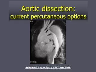
Diagnostic Imaging • Not performed until the patient is medically stable • Has been a dramatic shift from invasive to non-invasive diagnostic strategy • Spiral CT scan • TEE • MRI • Angiography
Imaging • Can identify aortic dissection and other features such as: • Involvement of the ascending aorta • Extent of dissection • Thrombus in the false lumen • Branch vessel or coronary artery involvement • Aortic insufficiency • Pericardial effusion with or without tamponade • Sites of entry and re-entry
Angiography • First definitive test for aortic dissection • Traditionally considered “the gold standard” • Involves injection of contrast media into the aorta • Identifies the site of the dissection • Major branches of the aorta • Communication site between true & false lumen • Can detect thrombus in the false lumen • Disadvantages • Not very practical in critically ill patients • Nephrotoxic contrast • Risks of an invasive procedure
Spiral CT • Sensitivity 83% • Specificity 90 - 100% • Two distinct lumens with a visible intimal flap can be identified • Advantages • Noninvasive • Readily available at most hospitals on an emergency basis • Can differentiate dissection from other causes of aortic widening (tumor, periaortic hematoma, fat) • Disadvantages • Sensitivity lower than TEE and MRI • Intimal flap is seen < 75% of cases • Nephrotoxic contrast is required • Cannot reliably detect AI, or delineate branch vessels
TTE • First used to diagnose aortic dissections in the ’70s • Sensitivity 59-85%, specificity 63-96% • Image quality limited by obesity, lung disease, and chest wall deformities
TEE • Sensitivity 98% Specificity 95% • Advantages • Close proximity of the esophagus to the thoracic aorta • Portable procedure • Yields diagnosis in < 5 minutes • Useful in patients too unstable for MRI • True and false lumens can be identified • Thrombosis, pericardial effusion, AI, and proximal coronary arteries can be readily visualized
TEE • Lower specificity attributed to reverberations atherosclerotic vessels or calcified aortic disease producing echo images that resemble an aortic flap • Disadvantages • Contraindicated in patients with esophageal varices, tumors, or strictures • Potential complications: bradycardia, hypotension, bronchospasm
MRI • Most accurate noninvasive for evaluating the thoracic aorta • Sensitivity 98% • Specificity 98% • Advantages • Safe • Can visualize the whole extent of the aorta in multiple planes • Ability to assess branch vessels, AI, and pericardial effusion • No contrast or radiation • Disadvantages • Not readily available on an emergency basis • Time consuming • Limited applicability in pts with pacemakers or metallic clips
Conclusions • Conventional TTE is of limited diagnostic value in assessment of the thoracic aorta • Both TEE and MRI have excellent sensitivity, however MRI is more specific • MRI is the study of choice for stable patients • TEE is the study of choice for unstable patients
Treatment • Acute dissections involving the ascending aorta are considered surgical emergencies • Dissections confined to the descending aorta are treated medically • Unless patient demonstrates continued hemorrhage into the pleural or retroperitoneal space
Surgical Options • Excision of the intimal tear • Obliteration of entry into the false lumen proximally • Reconstitution of the aorta with interposition of a synthetic vascular graft
Type A Dissections • Operative mortality varies from 7-35% • 27% post-op mortality • Patients who died had a higher rate of in-hospital complications such as strokes, renal failure, limb ischemia, & mesenteric ischemia
Poor prognostic factors • Hypotension or shock • Renal failure • Age> 70 yrs • Pulse deficit • Prior MI • Underlying pulmonary disease • Preoperative neurologic impairment • Renal and/or visceral ischemia • Abnormal EKG, particularly ST elevation
Medical therapy • Reduce systolic BP to 100 to 120 mmHg or the lowest level that is tolerated • IV Beta blockers • Propanolol (1-10 mg load, 3mg/hr) • Labetalol (20 mg bolus, 0.5 to 2 mg/min) • If SBP remains >100mmHg, nitroprusside should be added • Do not use without beta blockade • Avoid hydralazine • Surgical intervention for Type B dissections reserved for patients with a complicated course
Long Term Outcome • Type A • Survival at 5 yrs – 68% • Survival at 10 yrs – 52 % • Type B • 5 yrs – 60 - 80% • 10 yrs – 40 – 80% • Spontaneous healing of dissection is uncommon
Long-Term Management • Medical therapy • Oral Beta-blockers (reduces aortic wall stress) • Keep BP < 135/80 mmHg (combination therapy) • Avoidance of strenuous physical activity • Serial imaging • Thoracic MR scan prior to discharge • f/u scans at 3, 6, and 12 months • Subsequent screening studies done every 1-2 yrs if no evidence of progression