Pericarditis / Aortic Dissection
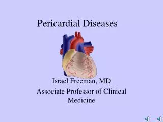
Pericarditis / Aortic Dissection. John G. McGinnity M.S., P.A.-C. Pericardium. Closed mesothelial sac Completely enveloping the heart from the base of the great vessels Inner serous membrane – visceral pericardium Outer fibrous layer – parietal pericardium

Pericarditis / Aortic Dissection
E N D
Presentation Transcript
Pericarditis / Aortic Dissection John G. McGinnity M.S., P.A.-C.
Pericardium • Closed mesothelial sac • Completely enveloping the heart from the base of the great vessels • Inner serous membrane – visceral pericardium • Outer fibrous layer – parietal pericardium • Ligamentous attachment to the sternum, vertebral column, and diaphragm
Pericardium • The layers are attached to each other by connective tissue with elastin fibers • Visceral pericardium produces 25-50ml clear fluid or more in case of disease • Drainage of the pericardial space occurs both by the thoracic via the parietal pericardium and by the right lymphatic duct via the right pleural space
Function of the Pericardium • Mechanical protection • Lubrication to reduce friction • Prevents spread of disease • Limit acute distension of the heart • Ligamentous attachments help to fix the heart in position
Abnormal Morphology • Congenital absence • Very rare
Acute Pericarditis • Syndrome due to inflammation of the pericardium characterized by chest pain, a pericardial friction rub, and serial electrocardiographic abnormalities.
Pericarditis Etiology • Viral Pericarditis – Coxsackie B Virus • Enteric viruses • Malignancy-lung, Breast CA and lymphoma are most common • Metabolic – Uremia up to 20% of patients on hemodialysis • Infection TB, SBE • Drug Induced – Procainamide, Hydralazine • Idiopathic Inflammatory disease – SLE, RA • Post Myocardial injury s/p AMI (1-3 weeks post)
Pericarditis Etiology • Radiation • Trauma • Fungal infection • Dissecting Aortic aneurysm • Idiopathic
Clinical Presentation • Chest pain is the most common presentation • Typical pain is retrosternal, radiation to left side of neck/back, trapezius, or scapula • Position dependent • Coughing or deep breathing increase chest pain • Leaning forward may give some relief to pain
Additional Symptoms • SOB / DOE may have shallow breath • Fever • Cough • Sputum production • Weight loss
Percordium / retrosternal Sharp / pleuritic Increased by breathing Hours / Days Leaning forward relief Retrosternal L Shd Pressure / burning Not related to breathing No effect on motion Angina pain only with exertion; USA rest pain associated Pericardial vs. Ischemic Pain
Viral syndrome Precursor • Recent viral syndrome -fever, cough, myalgias • Cough • Hiccups • Effusion: Dyspnea, tachycardia, edema, hypotension(late)
Physical Examination • Friction Rub – due to inflamed pericardial surfaces rubbing together; pericardial friction rub is pathognomonic for acute pericarditis • Relative tachycardia • Hypotension – Late • Usually self limiting • Watch out for Tamponade
Pulsus Paradoxus • Exaggerated response from the normal physiologic drop in BP with inspiration • Up to 10 mmHg drop in systolic BP occurs normally with inspiration • Tampanode>10mmHg drop • Total paradox complete disappearance of the palpable pulse - Severe
Laboratory • CBC, lytes, Bun/Cr. • Possible ANA, RF • PPD r/o Tb
ECG - Changes • Diffuse ST segment elevation with upright T waves • PR interval depression • Low voltage • A-fib /flutter
Treatment • Pain responds to NSAIDS, ASA • If no response 48 Hours – Steroids may be indicated consider Prednisone 60-80 mg q d in divided doses for 5-7 days then may tapered • Some patients can develop recurrent pericarditis and may need longer steroid therapy
Prognosis • Viral pericarditis idiopathic pericarditis,post-myocardial infarction pericarditis or post pericardiotomy syndrome are usually self limiting within 2-6 weeks • Sagrista-Sauleda et al. have observed by P.E. and noninvasive study that about 9 % develop signs mild cardiac constriction within 30 days.
Tamponade Pathophysiology • Inability to fill the cardiac chambers in diastole • Rising intra cardiac pressures • Reduction of stroke volume and cardiac output leads to hypotension
Beck’s Triad • Described in 1935, these 3 features are typical of Cardiac Tamponade • Hypotension • JVD (elevation in systemic venous pressure) • Muffled heart sounds
Echocardiogram in Tamponade • RA and ventricular collapse during diastole • As little as 15cc on fluid can be detected by 2D echo • Differentiate from other causes – Tumor compression or hematoma, RV infarct
Tamponade Treatment • Drainage of the fluid – Cath or echo guided • Treat symptoms as for pericarditis • Pericardiotomy or Window placement may be required • Biopsy has higher yield than fluid analysis
Aortic Dissection • At least 2000 cases per year • Early mortality as high as 1% per hour if untreated • Thought to be initiated by a tear in the intimal layer exposing an underlying diseased medial layer (creating a false lumen or by media hemorrhage • Most common for dissection to proceed antegrade (Around the arch) but can go retrograde (Back toward the arch)
Dissections • Majority of dissections occur in the ascending aorta within several centimeters of the valve (65%) • Descending aortic – distal to the left subclavian artery (20%) • Aortic arch (10%) • Abd Aorta (5%)
Classifications • DeBakey Classifications • Type I Originates in ascending aorta and propagates to the arch or beyond • Type II originate and is limited to the ascending aorta • Type III originate in the descending aorta and goes distal or rarely retrograde • Stanford • Type A All involving ascending aorta • Type B all others • Descriptive • Proximal /distal
Etiology • Peak incidence is in the 6-7th decades of life • Men 2:1 • H/O HTN 80% of cases • Bicuspid aortic valve • Marfans syndrome • Arteritis • Pregnancy 50% of dissection in women <40 • Eclampsia with High BPs • Trauma
Symptoms • Severe Chest pain 75-90% of cases • Sudden onset and severe at inception • Ripping/ tearing/ stabbing pain • Migration of the pain 70% of cases • Location anterior pain more common ascending involvement posterior pain descending involvement • Less common presentation- CHF, CVA, syncope, ischemic neuropathy, cardiac arrest or sudden death
P.E. • HTN or Hypotension • Pulse deficits • Murmur of AR • Neurological manifestations CVA, altered consciousness or coma
Diagnostic Findings • CXR – widened mediastinum or silhouette(80-90%) • Aortography – some surgeons may require • Contrast-enhanced CT (Spiral) • MRI- Thought to be the current “Gold Standard” (not ER STAT available) • TEE/TTE – TEE a close second
Management • Untreated mortality 25% at 24hrs, 50% at 1 wk, 75% at 1 month, >90%at 1 yr. • Monitor BP, rhythm, and urine output • 2 large bore IV’s • Arterial line • Central venous line in pt’s with hypotension or CHF
Management (Continued) • Reduce systolic blood pressure to 100-120 mmHg • Beta-blocker should be considered to reduce contractility • Morphine to treat CP • Surgical consultation