Download
1 / 16
220 likes | 328 Vues
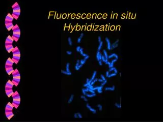
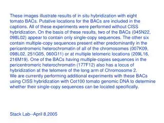
Fluorescence in situ hybridization (FISH), developed in 1980s, is a cytogenetic technique using fluorescent probes to bind the chromosome with a high degree of complementarity. It is a powerful and easy method to detect RNA or DNA sequences in cells, tissues and tumors. This technique is useful for identifying chromosomal abnormalities, gene mapping, characterizing somatic cell hybrids, checking amplified genes and studying the mechanism of rearrangements. RNA FISH is used to measure and localize mRNAs and other transcripts within tissue sections or whole mounts.<br><br>https://www.creative-bioarray.com/services/Fluorescent-In-Situ-hybridization-FISH.htm

E N D
WhatisFISH? Probes FISHProcedure Application 1 2 3 4 Definition, Principle and SampleTypes The core of FISH technology A quick and simple FISH protocol Wide range of uses and bright prospects Content
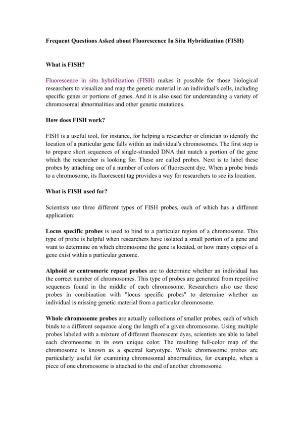
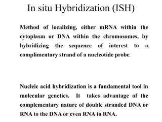
Definition • Fluorescent in situ hybridization (FISH) is a molecular cytogenetic technique that uses fluorescent probes that bind to only those parts of the chromosome with a high degree of sequence complementarity. • Itis used to detect and localize the presence or absence ofspecificDNAsequencesonchromosomes. What is Fluorescent in situ hybridization (FISH) ?
The principle of complementary base pairing The StructureofDNA The DNA contains two strand-like molecules coiled together into a structure known as a double helix. When two complementary sequences find each other they will bind together, or hybridise. • FISH works by exploiting the ability of one DNA strand to hybridise specifically to another DNA strand. HowdoesFISHwork ?
probe with fluorescent label heating cooling FISHTechnology hybridisation denaturation
Whole chromosome Centromere probes • Collection of probes that bind to the whole length of chromosome • Multiple probe labels are used • Hybridize along the length of the chromosome • Alpha and Satellite III probes • Generated from repetitive sequences found in centromeres • Centromere regions are stained brighter Probes Telomere Locus • WhatKindofProbesCanBeUsed? • Specific for telomeres • Specific to the 300 kb locus at the end ofspecific chromosome • Deletion • Translocation probes • Gene detection & localization probes • Gene amplification probes
dsDNA probes ssDNAprobes Stable, easier to work with, more specific, resistant to RNases, better tissue penetration, without self- hybridize Stable, available, easier to obtain Probes RNAprobes Synthetic oligonucleotides probes • CharacteristicsofDifferentProbesTypes • Higher thermal stability, • Better tissue penetration, • More specific, • Low background noise by RNase • Economical, stable, available, easier to work with, • more specific, resistant to RNases, • better tissue penetration, better reproducibility.
32P • 35S • 3H Radioactive isotopes Probes Labeling • Biotin • Digoxigenin • Fluorescent dye (FISH) • ProbesLabeling • Non-Radioactive isotopes
Preparation of the fluorescent probes • Hybridization of the probe and the target • Denaturation of the probe and the target Protocol Outline
FISHinCells The Sample Preparation and Sample Types • Sample Types • Fixed cell suspension • Formalin fixed paraffin embedded tissues • Preparation • Sample fixation • Slide tissue FISHintissues
Commercial Probes can be provided by many biotech companies • Based on your special needs, custom probes are also synthesized • e.g.𝒂-satellite DNA is often chosen as the source of centromeric probes. Preparation of the fluorescent probes
Dehydration(1) Place the suitable amount of fixed sample on the slide,(2) put into 46°C oven drying for 10 minutes(3) Followed by immersion 50%, 80%, 96% ethanol solution, each for 3 minutes(4) dry in the air Hybridization(1) Add 10 μl of Hybridization buffer to the sample on the glass plate and try to cover the entire sample(2) Add 1 μl probe to Hybridization buffer(3) Put a wet absorbent paper in a 50ml centrifuge tube, put the glass pieces in, then place them in a 46°C incubator for 1.5 hours(4) Preheat the Washing buffer to 48°C for the next step • Denaturation of the probe and the target
1 Morphology Morphology and population structure of microorganisms 2 Pathology Pathogen profiling, abnormal gene expression Developmental biology 3 Gene expression profiling in embryonic tissues Karyotyping and phylogenetic analysis • Applications Unique FISH patterns on individual chromosomes, chromosomal aberrations 4
01 • Products WeprovidecomprehensivecommercialprobesandFISHKitsforeasytouse. • CreativeBioarray • FISHServices 02 In addition to products, high-quality services are also available for our clients.
For more info please contact us: E-mail: info@creative-bioarray.com Gotoourwebsite: www. creative-bioarray.com