Download
1 / 48
540 likes | 1.99k Vues
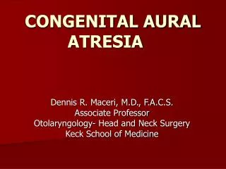
CONGENITAL AURAL ATRESIA. Dennis R. Maceri, M.D., F.A.C.S. Associate Professor Otolaryngology- Head and Neck Surgery Keck School of Medicine. “ The better developed the auricle the better the middle ear” K ountakis, 1995. Atresia. Epidemiology 1 in 10,000 to 15,000 births

E N D
CONGENITAL AURAL ATRESIA Dennis R. Maceri, M.D., F.A.C.S. Associate Professor Otolaryngology- Head and Neck Surgery Keck School of Medicine
“ The better developed the auricle the better the middle ear”Kountakis, 1995
Atresia Epidemiology • 1 in 10,000 to 15,000 births • Up to 50% of the time associated with some craniofacial syndrome • Unilateral : Bilateral, about 3:1 • 30% are bilateral • Atresia : Microtia, 7:1 • Slightly more common on the right • Male : Female, 2:1
Atresia Aural atresia associated with 22/71 known craniofacial syndromes • Treacher Collins (Mandibulofacial Dysostosis) • Nager Syndrome (Acrofacial Dysostosis) • Cruzoun’s Craniofacial Dysostosis • Goldenhar’s Syndrome • Hemifacial Microsomia
Aresia Surgery • First attempt to surgically correct aural atresia was by Thomson in 1843 • Shambaugh, 1967, recommended unilateral surgery only if the cochlear reserve allowed hearing to improve by 25dB • Jahrsdoerfer, 1978, first large series using the anterior approach
Embryology 8 Week Stage
Embryology 8 Week Stage
Embryology • The external ear forms earlier than the middle ear • Microtia and Atresia imply an arrest in development at any stage • The earlier the arrest, the less developed the middle ear
Embryology • Inner ear and labyrinth develop at 3 weeks from an invagination of ectoderm called the ottic placode • Middle ear space, mastoid, eustachian tube and part of the drum develop from the first pharyngeal pouch at 30 weeks • Mastoid air cells continue to develop up to age 5
Embryology • First Branchial Arch-(Meckel’s) • Malleus head, body, tensor tympani (V3) • Short process of the Incus • Second Arch (Hyoid) (Reicherts) • Manubrium of the malleus • Stapes superstructure • VIIth nerve • Foot plate from the otic capsule and 2nd arch
Embryology • External ear (ectoderm) is derived from the 1st branchial cleft. A solid core of epithelium migrates inward toward the 1st branchial pouch (endoderm)
Embryology Solid core of tissue migrates inward External canal re-canalizes by the 6th month
Embryology • The external canal starts to hollow out (recanalize) during the 6th month and progresses from medial to lateral • Arrest of recanalization process leads to the various deformities seen in atresia • Formed tympanic membrane and bony canal with a stenotic membranous canal leads to canal cholesteatoma
Embryology • Arrested growth of the ossicles leaves them in various stages of formation • Stapes often malformed but mobile • The incus and malleus are fused • Altered course of facial nerve
Atresia Embryology – 7th Month • Canalization complete • Mastoid separation from mandible Normal posterior-inferior growth No mastoid growth Normal Atresia
Atresia Embryology • Mastoid growth affects the facial nerve position Normal 120o Curve Acute Curve in Atresia
Classification of Deformities After Colman-3 types • Minor Aplasia-incomplete recanalization • Moderate Aplasia- the tympanic bone has developed but has failed to recanalize • Severe Aplasia-complete absence of the external canal
Atresia Classification-Severe Aplasia, no tympanic bone
Atresia Complete Atresia
Atresia Complete Atresia
Atresia Moderate Aplasia • The most common, solid mass of compact bone that has failed to recanalize
Atresia Minor Aplasia-partial recanalization • Middle space constricted, often with severe ossicular abnormalities
Microtia Types of Microtia (Based on Marx 1926) Class I Mild deformity, auricle smaller, all parts of ear identifiable Class II ½ to 2/3 normal size with partially retained structures Class III Severely malformed, peanut shaped
Radiological Evaluation • High resolution CT in coronal and axial planes • Axial to delineate malleus, incus and I-S joint and round window • Coronal to delineate stapes, oval window and vestibule • 3-D CT of little help • Timing indicated by timing of surgery
Grading System • Based on high resolution CT scan • A score of 5 or less denotes a poor candidate • Microtia indicates an arrest in development and abnormalities of the middle ear Kountakis, Helidonis and Jahrsdoerfer Arch. Oto 1995
Atresia Grade of Microtia as an Indicator of ME Development Microtia Grade Radiological Score I 8.5 II 7.2 III 5.9 Kountakis Arch Otolaryngol 1995
Radiological Evaluation • Good middle ear and mastoid aeration • Prominent ossicular mass with incus and malleus fused • Open oval window • Score of 8
Radiological Evaluation • Score of 8 2-stapes & OW 2-I-S joint, Incus 1-Aerated ME space 1-Bony cover on VII 1-Aerated mastoid 1-Pinna
Radiological Evaluation • Poor middle ear and mastoid development • No definable ossicular mass • Closed oval window • Not a good candidate • A score of 3
Audiological Evaluation • 50-60 dB conductive loss • Usually normal sensorineural function • Unilateral- behavioral audiometry • ABR for infants • Bilateral atresia presents a masking dilemma • Bone conduction ABR can help resolve the masking problem (Wave I on stimulated ear)
Surgical Considerations • Most consider repair in bilateral atresia • Many are reluctant to operate on unilateral cases • Not simply the hearing loss • Expectations of hearing recovery • Lifetime care of mastoid cavity • Potential risks to facial nerve and labyrinth • 55-65% achieve 25 dB speech-hearing level
Surgical Considerations • Most surgeons choose the anterior approach to avoid the mastoid cavity • 40% of patients with unilateral atresia are not surgical candidates such as those with severe aplasia as in Treacher Collins syndrome • Bilateral atresia- best ear by CT done as child approaches school age
Surgical Considerations Timing of surgery • Usually performed after age 6 or 7 years • This allows for microtia repair to be done first (If Medpor used,canalplasty done first) • Canal cholesteatoma in the stenotic ear usually develops in canals less than 2mm in diameter. If ear unfavorable, canalplasty alone is offered
Canal Cholesteatoma • Usually in ears with minor aplasia • Canal <4mm, 50% incidence
Surgical Technique • Minor aplasia- canal widening and middle ear ossicular work with tympanoplasty • Moderate Aplasia • Mastoid or posterior approach • Anterior approach
Surgical Technique Anterior Approach • Middle ear approached through the atretic bone with a limited mastoid opening
Surgical Technique • The posterior wall of the glenoid fossa becomes the anterior wall of the new ear canal • The epitympanum is the first part of the middle ear encountered • Fused ossicles identified
Surgical Technique • Atretic bone removed at times with a curette • Globular mass separated from the stapes to avoid cochlear trauma • Course of facial nerve determined • Ossiculoplasty performed • Tympanic membrane grafted • Meatoplasty • Split thickness skin graft (.008-.010 inches) lines the canal
Surgical Technique • The Meatoplasty must be aligned with the newly created bony canal
Surgical Technique • 4cm X 6cm split thickness skin graft • The graft is positioned in the canal and sewn to the meatal margin • Graft stabilized with Merocel wicks and hydrated with ear drops
Hearing Results • Post-op hearing level of 30 dB or better In 50-75% of patients with moderate or severe aplasia • 20 dB or better in 15-20% • Bellucci 20 dB in 50% @ 2 years • Schuknect similar results at 1.3 years • De La Cruz 56 patients 53% @ 20 dB at 6 mo. • Lambert early 60% @25 dB, 46% >1 yr.
Alternatives to Surgery • Bone anchored hearing aid (BAHA)
BAHA Hearing results better than with BC aids
Surgical Complications • Persistent or recurrent conductive hearing loss • Lateralization of graft • Scar tissue • SNHL • VIIth Nerve injury • 30 % revision rate • Re-stenosis • Graft migration • Inadequate hearing • Chronic cavity infection
Summary • Choose your patients carefully • Realize your potential v. patient expectations • Appropriate radiographic studies • Accurate audiological analysis • Facial nerve monitoring • Know when NOTto operate