Download
1 / 28
480 likes | 2.22k Vues
Chapter 3: DNA Replication Models of DNA replication: Meselson-Stahl Experiment DNA synthesis and elongation DNA polymerases Origin and initiation of DNA replication Prokaryote/eukaryote models (circular/linear chromosomes) Telomere replication

E N D
Chapter 3: DNA Replication • Models of DNA replication: Meselson-Stahl Experiment • DNA synthesis and elongation • DNA polymerases • Origin and initiation of DNA replication • Prokaryote/eukaryote models (circular/linear chromosomes) • Telomere replication
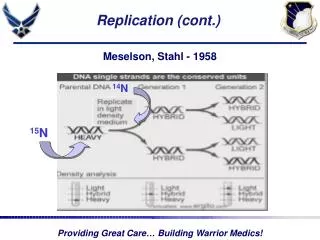
1958: Matthew Meselson & Frank Stahl’s Experiment Semiconservative model of DNA replication (Fig. 3.2)
1955: Arthur Kornberg • Worked with E. coli. • Discovered the mechanisms of DNA synthesis. • Four components are required: • dNTPs: dATP, dTTP, dGTP, dCTP (deoxyribonucleoside 5’-triphosphates) (sugar-base + 3 phosphates) 2. DNA template • DNA polymerase (Kornberg enzyme) 4. Mg 2+ (optimizes DNA polymerase activity) 1959: Arthur Kornberg (Stanford University) & Severo Ochoa (NYU)
Three main features of the DNA synthesis reaction: • DNA polymerase I catalyzes formation of phosphodiester bond between 3’-OH of the deoxyribose (on the last nucleotide) and the 5’-phosphate of the dNTP. • Energy for this reaction is derived from the release of two of the three phosphates. • DNA polymerase “finds” the correct complementary dNTP at each step in the lengthening process. • rate ≤ 800 dNTPs/second • low error rate 3. Direction of synthesis is 5’ to 3’ DNA polymerase Image credit: Protein Data Bank
There are many different types of DNA polymerase • Polymerase Polymerization (5’-3’) Exonuclease (3’-5’) Exonuclease (5’-3’) #Copies • I Yes Yes Yes 400 • II Yes Yes No ? • III Yes Yes No 10-20 • 3’ to 5’ exonuclease activity = ability to remove nucleotides from the • 3’ end of the chain • Important proofreading ability • Without proofreading error rate (mutation rate) is 1 x 10⁻⁶ • With proofreading error rate is 1 x 10⁻⁹ (1000-fold decrease) • 5’ to 3’ exonuclease activity functions in DNA replication & repair.
Eukaryotic enzymes: • Five DNA polymerases from mammals. • Polymerase (alpha): nuclear, DNA replication, no proofreading • Polymerase (beta): nuclear, DNA repair, no proofreading • Polymerase (gamma): mitochondria, DNA repl., proofreading • Polymerase (delta): nuclear, DNA replication, proofreading • Polymerase (epsilon): nuclear, DNA repair (?), proofreading • Different polymerases for nucleus and mtDNA • Some polymerases proofread; others do not. • Some polymerases used for replication; others for repair.
Origin of replication (e.g., the prokaryote example): • Begins with double-helix denaturing into single-strands thus exposing the bases. • Exposes a replication bubble from which replication proceeds in both directions. ~245 bp in E. coli
Initiation of replication, major elements: • Segments of single-stranded DNA are called template strands. • Gyrase (a type of topoisomerase) relaxes the supercoiled DNA. • Initiator proteins and DNA helicase binds to the DNA at the replication fork and untwist the DNA using energy derived from ATP (adenosine triphosphate). (Hydrolysis of ATP causes a shape change in DNA helicase) • DNA primase next binds to helicase producing a complex called a primosome (primase is required for synthesis), • Primase synthesizes a short RNA primer of 10-12 nucleotides, to which DNA polymerase III adds nucleotides. • Polymerase III adds nucleotides 5’ to 3’ on both strands beginning at the RNA primer. • The RNA primer is removed and replaced with DNA by polymerase I, and the gap is sealed with DNA ligase. • Single-stranded DNA-binding (SSB) proteins (>200) stabilize the single-stranded template DNA during the process.
DNA replication is continuous on the leading strand and semidiscontinuous on the lagging strand: • Unwinding of any single DNA replication fork proceeds in one direction. • The two DNA strands are of opposite polarity, and DNA polymerases only synthesize DNA 5’ to 3’. • Solution: DNA is made in opposite directions on each template. • Leading strand synthesized 5’ to 3’ in the direction of the replication fork movement. • continuous • requires a single RNA primer • Lagging strand synthesized 5’ to 3’ in the opposite direction. • semidiscontinuous (i.e., not continuous) • requires many RNA primers
Supercoiled DNA relaxed by gyrase & unwound by helicase + proteins: 5’ SSB Proteins Okazaki Fragments ATP 1 Polymerase III 2 Helicase + Initiator Proteins 3 Lagging strand 3’ primase base pairs 5’ Polymerase III RNA primer replaced by polymerase I & gap is sealed by ligase 5’ 3’ Leading strand RNA Primer 3’
DNA ligase seals the gaps between Okazaki fragments with a phosphodiester bond (Fig. 3.7)
Fig. 3.5 - Model of DNA replication Peter J. Russell, iGenetics: Copyright © Pearson Education, Inc., publishing as Benjamin Cummings.
Fig. 3.5 - Model of DNA replication Peter J. Russell, iGenetics: Copyright © Pearson Education, Inc., publishing as Benjamin Cummings.
Concepts and terms to understand: Why are gyrase and helicase required? The difference between a template and a primer? The difference between primase and polymerase? What is a replication fork and how many are there? Why are single-stranded binding (SSB) proteins required? How does synthesis differ on leading strand and lagging strand? Which is continuous and semi-discontinuous? What are Okazaki fragments?
Replication of circular DNA in E. coli (3.10): Two replication forks result in a theta-like () structure. As strands separate, positive supercoils form elsewhere in the molecule. Topoisomerases relieve tensions in the supercoils, allowing the DNA to continue to separate.
Rolling circle model of DNA replication(3.11): Common in several bacteriophages including . Begins with a nick at the origin of replication. 5’ end of the molecule is displaced and acts as primer for DNA synthesis. Can result in a DNA molecule many multiples of the genome length (and make multiple copies quickly). During viral assembly the DNA is cut into individual viral chromosomes.
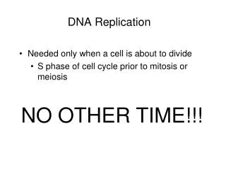
DNA replication in eukaryotes: Copying each eukaryotic chromosome during the S phase of the cell cycle presents some challenges: Major checkpoints in the system Cells must be large enough, and the environment favorable. Cell will not enter the mitotic phase unless all the DNA has replicated. Chromosomes also must be attached to the mitotic spindle for mitosis to complete. Checkpoints in the system include proteins call cyclins and enzymes called cyclin-dependent kinases (Cdks).
Each eukaryotic chromosome is one linear DNA double helix • Average ~108 base pairs long • With a replication rate of 2 kb/minute, replicating one human chromosome would require ~35 days. • Solution ---> DNA replication initiates at many different sites simultaneously. Rates are cell specific! Fig. 3.14
What about the ends (or telomeres) of linear chromosomes? DNA polymerase/ligase cannot fill gap at end of chromosome after RNA primer is removed. this gap is not filled, chromosomes would become shorter each round of replication! Solution: Eukaryotes have tandemly repeated sequences at the ends of their chromosomes. Telomerase (composed of protein and RNA complementary to the telomere repeat) binds to the terminal telomere repeat and catalyzes the addition of of new repeats. Compensates by lengthening the chromosome. Absence or mutation of telomerase activity results in chromosome shortening and limited cell division.
Fig. 3.16 Synthesis of telomeric DNA by telomerase Peter J. Russell, iGenetics: Copyright © Pearson Education, Inc., publishing as Benjamin Cummings.
Final Step - Assembly into Nucleosomes: • As DNA unwinds, nucleosomes must disassemble. • Histones and the associated chromatin proteins must be duplicated by new protein synthesis. • Newly replicated DNA is assembled into nucleosomes almost immediately. • Histone chaperone proteins control the assembly. Fig. 3.17