HEPATOBILIARY IMAGING
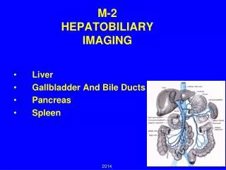
HEPATOBILIARY IMAGING. Presented by Yang Shiow-wen 11/26/2001. Hepatobiliary Imaging. Evaluates hepatocellular function and patency of the biliary system Tracing the production and flow of bile from the liver through the biliary system into the small intestine

HEPATOBILIARY IMAGING
E N D
Presentation Transcript
HEPATOBILIARY IMAGING Presented by Yang Shiow-wen 11/26/2001
Hepatobiliary Imaging • Evaluates hepatocellular function and patency of the biliary system • Tracing the production and flow of bile from the liver through the biliary system into the small intestine • Sequential images of the liver, biliary tree and gut are obtained • A "HIDA" scan or a "DISIDA" scan
Hepatobiliary Imaging • Performed with a variety of compounds that share the common imminodiacetate moiety
Structures of IDA derivates • Blue color: A polar component (the diacetate) • Red: A lipophilic component
IDA-chelated Tc-99m • A magnification of two imminodiacetate compounds • Polar components chelated a Tc-99m molecule
Pathways of IDA derivates • Thelipophilic component : binding to hepatocyte receptors for bilirubin • Transported through the same pathways as bilirubin, except for conjugation • Excretion decreased with increasing bilirubin levels
HIDA • HIDA • Little used today
DISIDA (Disofenin) • 85% extracted by the hepatocytes • Visualization of gallbladder and CBD when bilirubin > 8 ng/dl
BRIDA (Mebrofenin) • 98% extracted by the hepatocytes(bilirubin <1.5 mg/dL) • Visualization of gallbladder and CBD when bilirubin > 30 ng/dl • Higher hepatic extraction
BRIDA (Mebrofenin) • Rapid biliary to bowel transit time • Taken into consideration when evaluating acute cholecystitis • Mebrofenin may be preferred over Disofenin in suspected biliary atresia
Indications • Functional assessment of the hepatobiliary system • Integrity of the hepatobiliary tree • Evaluation of suspected acute cholecystitis • Evaluation of suspected chronic biliary tract disorders • Evaluation of common bile duct obstruction • Detection of bile extravasation • Evaluation of congenital abnormalities of the biliary tree
Contraindications • Hypersensitivity to • IDA derivative • Local anesthetics of the amide type • With disturbances of cardiac rhythm or conduction • Pregnancy Category: C
Requirements for DISIDA Scan • Patient preparation: fasted for 2-4 hours • Otherwise delayed or non-visualization • Fasted for > 24 hrs or on TPN, a false-positive study may occur • Radiotracer • Adult • 1.5-5 mCi Tc-99m IDA compounds i.v. • 3 – 10 mCi for hyperbilirubinemia • Children • 0.05 – 0.2 mCi/kg • minimum of 0.3 – 0.5 mCi
Requirements for DISIDA Scan Additional information • History of previous surgeries, especially biliary and gastrointestinal • Time of most recent meal • Current medications • esp. opioid compounds • Delaying the study for 4 hr after the last dose • Bilirubin and liver enzyme levels • Results of ultrasound
Requirements for DISIDA Scan • Gamma camera • A large field of view with a low energy all purpose or high resolution collimator • A smaller field of view with a diverging collimator
Requirements for DISIDA Scan • Serial anterior views for 60 minutes • Until activity is seen in both the gallbladder (patency of the cystic duct) and the small bowel (patency of the common bile duct) • Every 5 minutes for 30 minutes • Once at 45 minutes • Once at 1 hour • Right lateral views • At 30, 60 minutes • Oblique views • Separate gallbladder from small bowel activity • Delayed views • At 2 hours, 4 hours, 6 hours or 24 hours after injection • Severely ill patient, suspected CBD obstruction, suspected biliary atresia
Interventions • CCK (0.01-0.02 ug/kg) • Fasting for >24-48 hours, or on TPN • Empty the gall bladder (low resistance to bile flow state) • Preferential gallbladder filling • Delayed biliary to bowel transit • Injection 30 min prior to the test • Administered slowly (3 – 5 min) • Prevent biliary spasm and abdominal cramps • Water (5-10 cc) • Distinguish transient duodenal activity from gallbladder
Interventions • Morphine sulfate (0.04-0.1 mg/kg) • When acute cholecystitis is suspected and the GB is not seen by 60 min • & Radiotracer within the small intestine • Enhancing sphincter of Oddi tone • Increasing pressure within the CBD • Diverting bile away from the sphincter of Oddi & into functionally obstructed sludge filled gallbladder
Interventions • Fatty meal stimulation • Gallbladder ejection fraction measurement • Phenobarbital • When biliary atresia is suspected • 5 mg/kg/day (orally) for 3 – 5 days prior to the study • Enhancing the biliary excretion of the radiotracer
Processing • Gallbladder ejection fraction (GBEF) • Using the immediate pre-CCK and the post-CCK data • Regions of interest (ROI) are drawn around the GB and adjacent liver (background) • Hepatic extraction fraction (HEF) • Index of hepatocellular function • Deconvolution analysis from ROI over the liver and heart
Normal Study • Immediate demonstration of hepatic parenchyma • Prompt clearance of the blood pool within the first 5 minutes • Biliary excretion should commence within 20 minutes (5-10 min) • Biliary ducts would visualize followed the gallbladder • Gallbladder and small bowels are visualized within 1 hour
Acute Cholecystitis • The most common indication • S\S • Nausea, vomiting, fever • Right upper quadrant pain post-prandially • Mild to moderate leukocytosis • Abnormal liver function test • Pain radiates to the back (scapula) • Obstruction of cystic duct • By a gallstone • Inflammation, edema, gallbladder mucous, or a tumor (5%)
Acute Cholecystitis • DISIDA scan • Sensitivity: 95%, specificity 93-96% • Positive predictive value: 92.1%, negative predictive value: 99% • Adequate filling of the gallbladder • Acute cholecystitis is effectively excluded • Cystic duct obstruction • Failure to visualize the gallbladder up to 4 hours
Acute Cholecystitis • When acute cholecystitis is suspected and the gallbladder is not seen within 40–60 min • 3 – 4 hr delayed images should be obtained • Rule out chronic cholecystitis • Premedication with CCK • Morphine augmentation
Acute Cholecystitis • Premedication with CCK • Same sensitivity and specificity • Disadvantages • Not differentiated chronic cholecystitis from normal • Nausea, vomiting, exacerbation of bladder pain • Missed acute cholecystitis exhibiting delayed gallbladder visualization • Without delayed views • Malrotaion, enterogastric reflux, masses displacing or inflammatory processes of the small bowel
Acute Cholecystitis • Ingestion of morphine sulfate • More accurately, less complication • Differential diagnosis for non-visualization of the gallbladder • Relaxation of the sphincter of Oddi • Imaging is usually continued for another 30 min • Contraindications • Absolute: Respiratory depression in non-ventilated patients, morphine allergy • Relative: acute pancreatitis
Acute Cholecystitis • The hallmark of acute cholecystitis (acalculous as well as calculous) • Persistent gallbladder non-visualization 30 min post-morphine or on the 3 – 4 hr delayed image • Rim sign • A band or rim of increased activity adjacent to gallbladder fossa • Associated with severe phlegmonous/gangrenous acute cholecystitis, a surgical emergency • Cystic duct obstruction, acute cholecystitis
Chronic Cholecystitis • Ultrasound is the primary modality of choice • S\S • Usually having gall stones • The cystic duct is not blocked • More chronic pain
Common Bile DuctObstruction • Delayed visualization of the gall bladder • Clinical settings associated with physiologic failure of the gallbladder to filling • e.g. fasting for >24 – 48 hr, severely ill or post-operative patients may result in GB non-visualization within the first hour • A larger dose of morphine (0.1 mg/kg) decrease the false positive rate • Separated from acute cholecystitis using morphine or delayed imaging • Reduced gallbladder ejection fraction in response to CCK • Indicative of chronic cholecystitis, gallbladder dyskinesia or the cystic duct syndrome • Visualization of the GB after the bowel
Common Bile Duct Obstruction • S\S • Hyperbilirubinemia (> 5 mg/dl) • Dilation of CBD (sonography, >3 days) • A history of pancreatitis (serum amylase) • DISIDA scan • High grade or a total CBD obstruction • Sensitivity: 95% • Detection immediately
Common Bile Duct Obstruction • Delayed biliary-to-bowel transit beyond 60 min raises the suspicion • Activity in the small bowel seen within 60 min does not entirely exclude partial CBD obstruction • When neither the gallbladder nor the small bowel are seen within 18–24 hrs • Suspected High grade CBD obstruction • Severe hepatocellular dysfunction may appear similar
Bile Leaks • Most appropriate non-invasive imaging technique for evaluation of bile leaks • Sensitivity: 87%, Specificity: 100% (2-3 ml of labeled bile) • Radiopharmaceutical activity • In an extrahepatic and extraluminal location • More intense with time • Differentiating intraluminal activity from a leak • Standing views in addition to decubitus views • Cinematic display • 3 – 4 hrs delayed imaging
Biliary Atresia • Excluded by demonstrating transit of radiotracer into the bowel • Failure of tracer to enter the gut • Hepatocellular disease • Immature intrahepatic transport mechanisms • Biliary atresia • CBD obstruction • Urinary excretion of the tracer (especially in diaper) may be confused with bowel activity
Duodenogastric Bile Reflux • Highly correlated with bile gastritis • Cause of epigastric discomfort
False Positive Study • Gallbladder non-visualization in the absence of acute cholecystitis • Insufficient fasting (<2 – 4 hr) • Prolonged fasting (>24 – 48 hr), especially total parenteral nutrition (despite CCK pre-treatment and Morphine augmentation) • Severe hepatocellular disease • High grade common bile duct obstruction • Severe intercurrent illness (despite CCK pre-treatment and Morphine augmentation) • Pancreatitis (rare) • Rapid biliary-to-bowel transit (insufficient tracer activity remaining in the liver for delayed imaging) • Severe chronic cholecystitis • Previous cholecystectomy
False Negative Study • Gallbladder visualization in the presence of acute cholecystitis • Bowel loop simulating gallbladder (drinking water may help to clarify anatomy) • Acute acalculous cholecystitis • The presence of the "dilated cystic duct" sign simulating GB. (Morphine should not be given) • Bile leak due to GB perforation • Congenital anomalies simulating gallbladder • Activity in the kidneys simulating gallbladder or small bowel (may be clarified by a lateral image)
References • http://www.vh.org/Providers/Lectures/IROCH/BiliaryNucs/BiliaryNucs.html (Virtual Hospital) • http://www.cancerboard.ab.ca/about/ercdocs/diiso.html • http://www.nuclearonline.org/PI/Bracco%20mebrofenin%20doc.pdf • http://www.snm.org/pdf/hb2.pdf • http://www.vh.org/Providers/Textbooks/ElectricGiNucs/Text/Hepatobiliary.html • Chapter 38, Hepatobiliary Imaging, Darlene Fink-Bennett, P759-770
The End Thank for Your Attention !