Download
1 / 71
980 likes | 1.89k Vues
ABO and H Blood Groups. Terry Kotrla, MS, MT(ASCP)BB 2010. History. Discovered in 1900 by Karl Landsteiner and remains the most important blood group system Mixed blood of colleagues (serum from one, cells from another) together Discovered A, B and O His student discovered AB in 1902.

E N D
ABO and H Blood Groups Terry Kotrla, MS, MT(ASCP)BB 2010
History • Discovered in 1900 by Karl Landsteiner and remains the most important blood group system • Mixed blood of colleagues (serum from one, cells from another) together • Discovered A, B and O • His student discovered AB in 1902
Landsteiner’s Rule • Reciprocal antibodies are consistently and predictably present in the sera of normal people whose rbcs lack the corresponding antigen(s) • He was awarded the Nobel Prize in Physiology or Medicine in 1930 for his work
Red Cell membrane structure • Surface of the RBC consists of a bilipid membrane in which large protein molecules are embedded. • Composed of phosholipids which are both hydrophilic (heads) and hydrophobic (tails).
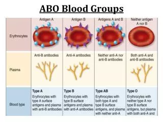
Red Blood Cell Membrane • External surface of RBC membrane is coated with a diverse array of glycoproteins, complex carbohydrates, and lipoproteins, imparting antigenic structure to the membrane.
A and B Antigens • Inheritance follows Mendelian genetics • Frequency in white population: group O 45%, group A 40%, group B 11% and group AB 4%. • Frequencies differ in selected populations and ethnic groups • Group B higher in Black and Asian populations
A and B Antigens • Not fully developed at birth, fewer copies of the antigen on the cells. • Antigens detectable as early as 5 weeks after conception • Human typing sera may give weaker, or very rarely negative, reactions
Biochemistry of A, B, H Antigens • ABO antigens are located on RBCs, lymphs, platelets, tissue cells, bone marrow and solid organs. • Inheritance results in expression. • Antigens ARE NOT direct products of the genes. • Genes code for TRANSFERASE which causes transfer of monosaccharide molecule onto a precursor substance on the RBC.
H Gene • Codes for the production of fucosyl transferase that catalyzes the addition of L-fucose,the immunodominant structure of H antigen. • Two slightly different structures, known as the type 1 and type 2 precursor chains. • The H gene and its allele h are inherited independently of the allelic A, B and O genes • H antigen only person is group O. • Once L-fucose added A and B gene specified products can be add their sugars.
A Gene • Codes for production of a galactosaminyl transferase that effects the addition of N-acetyl-galactosamine to the preformed H-bearing chains.
B Gene • Codes for production of a galactosyl transferase that effects the addition of D-galactose to the same H-bearing structure.
A, B and H • Immunodominant structure of the H antigen is L-fucose – person is group O • Immunodominant structure of A antigen N-acetyl-galactosamine – person is group A • Immunodominant structure of the B antigen, D-galactose- person is group B
Bombay (Oh) • If L-fucose is added H, A and B cannot be added. • Individual may inherit A or B genes but without H the immunodominant sugars CANNOT be added. • Must be homozygous for h, which is an amorph, with no detectable product, similar to d. • A Bombay individual has genotype hh but WILL type as Group O. • Can only be determined by family studies or testing with lectins
The H System • Two genes: H and h • H 99.9%> • H <0.% • Leads to production of H antigen – precursor molecule for A and B antigens. • Ulex europaeus = anti-H • Not all H is converted to A or B, varies among the blood groups. • Memorize: 0>A2>B>A2B>A1>A1B
H Antibodies • H-like antigens are found in nature. • Anti-H occasionally detected in A1, A1B and rarely in B individuals and will cause false positive in the REVERSE reaction. • So little H on cells may form anti-H • Relatively weak • Almost always reacts at RT • Considered clinically insignificant
H Antibodies • May cause an ABO discrepancy
H Antibodies • In contrast persons of the rare Oh (hh) phenotype (have no A, B or H antigens on their RBCs) form a potent clinically significant anti H which reacts well over a wide thermal range and with all RBCs except those of other O people.
Oh Phenotype (Bombay) • Occurs when two hh genes are inherited at the Hh locus. • Possess normal A or B genes (if they were inherited) but unable to express. • Must have H on red cell membrane • Can transmit A or B gene to offspring • Term “Bombay” used since first discovered in Bombay, India
Oh Phenotype (Bombay) • Symbol “Oh” denotes this phenotype • RBCs not agglutinated by anti-A, -B or –A,B • Serum/plasma agglutinates A and B cells • Not recognized until serum tested against group O cells and causes strong agglutination. • Have anti-A, -B, -A,B and –H • Can only be transfused with Bombay blood <0.01%
Oh Phenotype (Bombay) • Confirmatory testing • Anti-H lectin (Ulex europaeus) – negative • Agglutination of A, B, AB and O cells • Serum/plasma will not agglutinate Oh cells.
Secretor Genes • A, B and H antigens may be present in fluids. • Controlled by Se and se, secretor genes. • Need only one copy of the Se gene. • The gene se is an amorph. • Not linked to ABO locus, inherited independently
Secretor Genes • Persons who have A, B and/or H in secretions are called “secretors”
Secretor Genes • Secretor studies helpful in defining weak subgroups or resolving genetic make up of individuals with unusual blood group • 80% of Caucasians are secretors • 20% are non-secretors
Subgroups of A (A1 and A2) • Subgroups of A are phenotypes that differ from others of the same ABO group with respect to the • amount of A antigen carried on RBCs, and, • in secretors, present in the saliva. • Variant gene produces a weaker than normal red cell antigen
Subgroups of A (A1 and A2) • Different levels of expression of A (or B) on RBCs are classified into subgroups • 80% of group A individuals are A1 • Approximately 20% are A2 • Transferase produced by A2 gene differs from that produced by A1, less efficient in converting H chains to A
Difference Between A1 and A2 • A1 has more A and less H antigen on the cell. • A2 has less A and more H antigen • Cannot be detected serologically • A2 can produce anti- A2 – qualitative difference?
Lectins • Naturally occurring materials (usually plant) that react specifically with blood group antigens. • Dolichos biflorus – anti-A1 • Will agglutinate A1 red blood cells • Will not agglutinate A2 red blood cells
Anti-A1 • 1-8% of A2 and 22-35% of A2B people will have anti-A1 • Causes ABO discrepancy – reverse type • Incompatible crossmatch if donor A1 • NOT clinically significant unless reactive at 37C or AHG. • Clinically significant – ability to cause red cell destruction – donor blood or hemolytic disease of the fetus and newborn.
Subgroups of A weaker than A2 • Occur infrequently, characterized by decreasing numbers of A antigens • Less than 1% of total A gene pool
Classification of Weak A Subgroups • Strength of agglutination when tested with: • Anti-A1 lectin • Anti-A,B • Anti-H lectin • Presence of anti-A1 • Presence of A and H in secretions
Subgroups of A • Subgroups of A weaker than A2 (Ael, Aint, A3, Ax, Am, etc) are seen only infrequently • NOTE: A3 is characterized by mixed field agglutination.
Subgroups of B • Less common than subgroups of A • Criteria resembles that used for A subgroups • Usually detected based on forward type, reverse type correctly • Do not make anti-B as commonly • NOTE: B3 characterized by mixed field agglutination
Antibodies to A and B • Landsteiner – individuals do not form antibodies against antigens on their own cells. • “Naturally ocurring” is a misnomer • Antibody production stimulated by substances in the environment similar to blood group antigens • Simply implies antibody production NOT due to actual exposure to red blood cells • Allows both serum/plasma and red cells to determine ABO type – check/balance system
Development of anti-A and anti-B • Antibody production first few months of life • Babies cannot be reversed typed: • Antibodies present in baby from mom • Are not born with antibodies, detectable at 3 to 6 months of age • Once produced remain constant until elderly • Complete absence of ABO antibodies exceedingly rare
Anti-A,B • Group O have THREE ABO antibodies: anti-A, -B and –A,B • may react more strongly than anti-A and anti-B with some weak A or B subgroups • use anti-A,B to test group O donors • used to type babies, antigens not being well-developed at birth
Antibody Characteristics • React best at room temperature. • Agglutinate saline suspended red cells, no additional reagents are necessary • May produce hemolysis in vivo and in vitro
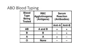
Routine Testing For ABO Group • Forward -Test known anti-serum with unknown patient cells (antigens) • Reverse – Test unknown serum (antibody) with known A and B antigens (cells0 • MUST MATCH
ABO Discrepancies • Discrepancy between forward and reverse • Helpful observations • Strength of reaction: forward 4+ if antigen present, reverse 2-4+. • Unexpected negative in reverse • Unexpected positive in forward OR reverse • Must delay transfusion until resolved – emergency give out O RBCs and appropriate D type
ABO Discrepancies • Errors divided into two categories • Technical • Sample • Results in false positives and negatives • Sample errors divided into rbc and serum
ABO Discrepancy – Technical False Negative • Failure to add serum or antiserum to a test. • Failure to identify hemolysis as a positive reaction. • Not using the appropriate serum (or reagent) to cell ratio. • Improper centrifugation. • Incubation of tests at temperatures above 20-25 C. • Use of inactive reagents. • Failure to interpret or record test results correctly.
ABO Discrepancy – Technical False Positive • Over centrifugation • Use of contaminated reagent antibodies, RBCs or saline • Use of dirty glassware • Incorrect interpretation or recording of results
Problems Associated With Testing RBC • Sample from recently transfused or bone marrow transplant patient • ABO subgroup or weakened antigens due to disease such as leukemia • Abnormal proteins or Wharton’s jelly • Increased A or B blood group substances inhibit reaction – false negative • Antibodies to dyes in forward reagents • Cold autoagglutinins – patient cells may spontaneously agglutinate