BENIGN EYELID LESIONS
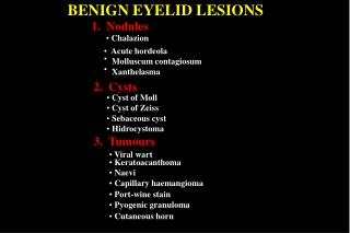
BENIGN EYELID LESIONS. 1. Nodules. Chalazion . Acute hordeola. Molluscum contagiosum. Xanthelasma. 2. Cysts. Cyst of Moll Cyst of Zeiss Sebaceous cyst Hidrocystoma. 3. Tumours. Viral wart. Keratoacanthoma. Naevi. Capillary haemangioma. Port-wine stain.

BENIGN EYELID LESIONS
E N D
Presentation Transcript
BENIGN EYELID LESIONS 1. Nodules • Chalazion • Acute hordeola Molluscum contagiosum Xanthelasma 2. Cysts • Cyst of Moll • Cyst of Zeiss • Sebaceous cyst • Hidrocystoma 3. Tumours • Viral wart • Keratoacanthoma • Naevi • Capillary haemangioma • Port-wine stain • Pyogenic granuloma • Cutaneous horn
Signs of chalazion (meibomian cyst) Painless, roundish, firm lesion within tarsal plate May rupture through conjunctiva and cause granuloma
Histology of chalazion Epithelioid Multinucleated cells giant cells Multiple, round spaces previously containing fat with surrounding granulomatous inflammation
Injection of local anaesthetic Treatment of chalazion Incision and curettage Insertion of clamp
Acute hordeola Internal hordeolum ( acute chalazion ) External hordeolum (stye) • Staph. abscess of meibomian • glands • Staph. abscess of lash follicle and • associated gland of Zeis or Moll • Tender swelling at lid margin • Tender swelling within tarsal plate • May discharge through skin • May discharge through skin • or conjunctiva
Molluscum contagiosum Signs Complications • Painless, waxy, umbilicated nodule • Chronic follicular conjunctivitis • May be multiple in AIDS patients • Occasionally superficial keratitis
Histology of molluscum contagiosum • Lobules of hyperplastic epithelium • Intracytoplasmic (Henderson-Patterson) • inclusion bodies • Circumscribed lesion • Surface covered by normal • epithelium except in centre • Deep within lesion bodies are small and • eosinophilic • Near surface bodies are larger and • basophilic
Xanthelasma • Common in elderly or those with • hypercholesterolaemia • Yellowish, subcutaneous plaques • containing cholesterol and lipid • Usually bilateral and located medially
Eyelid cysts Eccrine sweat gland hidrocystoma Cyst of Moll • Translucent • On anterior lid • margin • Similar to cyst of Moll • Not confined to lid • margin Cyst of Zeis Sebaceous cyst • Cheesy contents • Frequently at • inner canthus • Opaque • On anterior lid • margin
Viral wart (squamous cell papilloma) • Most common benign lid tumour • Raspberry-like surface Pedunculated Sessile
Histology of viral wart Finger-like projections of fibrovascular connective tissue Epidermis shows acanthosis (increased thickness) and hyperkeratosis Rete ridges are elongated and bent inwards
Keratoses Seborrhoeic Actinic • Common in elderly • Affects elderly, fair-skinned individuals • Discrete, greasy, brown lesion • Most common pre-malignant skin lesion • Friable verrucous surface • Rare on eyelids • Flat ‘stuck-on’ appearance • Flat, scaly, hyperkeratotic lesion
Keratoacanthoma • Lesion above surface epithelium • Uncommon, fast growing nodule • Acquires rolled edges and keratin-filled • crater • Central keratin-filled crater • Involutes spontaneously within 1 year • Chronic inflammatory cellular infiltration • of dermis
Naevi • Appearance and classification determined by location within skin • Tend to become more pigmented at puberty Intradermal Junctional Compound • Elevated • Flat, well-circumscribed • Has both intradermal • and junctional • components • May be non-pigmented • Pigmented • Low malignant potential • No malignant potential
Capillary haemangioma • Rare tumour which presents soon after birth • May be associated with intraorbital • extension • Starts as small, red lesion, most frequently • on upper lid • Grows quickly during first year • Begins to involute spontaneously • during second year • Blanches with pressure and swells on crying
Periocular haemangioma Treatment options • Steroid injection in • most cases • Surgical resection in • selected cases Occasional systemic associations • High-out heart failure • Kasabach-Merritt syndrome - • thrombocytopenia, anaemia • and reduced coagulant factors • Maffuci syndrome - skin • haemangiomas, • endrochondromas and • bowing of long bones
Histology of capillary haemangioma Lobules under high magnification Lobules of capillaries Fine fibrous septae
Port-wine stain (naevus flammeus) • Rare, congenital subcutaneous lesion • Segmental and usually unilateral • Does not blanch with pressure Associations • Ipsilateral glaucoma in 30% • Sturge-Weber or • Klippel-Trenaunay-Weber • syndrome in 5%
Progression of port-wine stain Initially red and flat Subsequent darkening and hypertrophy of skin Skin becomes coarse, nodular and friable
Pyogenic granuloma Cutaneous horn • Usually antedated by surgery or trauma • Uncommon, horn-like lesion protruding • through skin • Fast-growing pinkish, pedunculated or • sessile mass • May be associated with underlying actinic • keratosis or squamous cell carcinoma • Bleeds easily