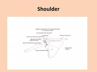
Shoulder lesions
Shoulder lesions. H.Makhmalbaf MD Consultant Orthopedic & Knee surgeon Ghaem Hospital Medical School. Painful arc syndrome . Pain in mid ROM of abduction Partial supraspinatus tendon tear Tendinitis of Supraspinatus Calcium deposite in supraspin. Tendon Sub-acromial bursitis


Shoulder lesions
E N D
Presentation Transcript
Shoulder lesions H.Makhmalbaf MD Consultant Orthopedic & Knee surgeon Ghaem Hospital Medical School
Painful arc syndrome • Pain in mid ROM of abduction • Partial supraspinatus tendon tear • Tendinitis of Supraspinatus • Calcium deposite in supraspin. Tendon • Sub-acromial bursitis • Greater tuberosity fracture
Painful arc syndrome • Pain in 45° abduction – 160 • X-ray calcium deposit or # • H/o trauma? Or inflamation • Conservative treatment mainly • Or acromioplasty
Frozen shoulder • Painful restriction of ROM • Unknown etiology • Gradually progressive • Chronicity • Slow spontaneous restoration of ROM
Etiology • Tendinitis of rotator cuff • Bicipitaltenosynovitis • Muscle imbalance, inactivity • RSD • Association with cardiovascular dis. • Trauma; degeneration; granulation tissue; adhesions
Clinical picture • 5th & 6th decades particularly women • Insidious onset ;injury ? Inactivity • Pain over anterolateral of shoulder • Worse at night • Limitation of active & passive ROM • Muscle spasm; internal rotation • Tenderness over bicipital grove
Treatment: • Bed rest , heat , sedation • If RA :injection of steroids & oral • Pendulum exercises • No forcible movement • Only MUA if necessary • Rarely need surgical release
Lesions of the rotator cuff • Aging, degenerative change • Rupture of cuff, deltoid takes over • Recurring pain & stiffness, aggravated by activity in the shoulder & arm • Tenderness over tuberosity & bicipital grove • H/o fall or lifting, acute pain & snap
Rotator cuff rupture • Unable to abduct arm • 45’ abd. by deltoid, 45- 90 is painful • Size of tear is important • Repair acute tear • Conservative neglected & old
Calcified deposits in the rotator cuff • Ca phosphate & Ca carbonate • In the tendon, lig. & capsule • Mainly in the white collar middle age • Freq. Bilateral, men 3rd& 4th decades • Gradual or acute, pain, lim. Abduction • X-ray Ca deposits
Treatment : • Conservative : • Ice bag, rest , needle aspiration irrigation • Diathermy, steroids, exercises • Surgical : • Relieves pain completely • Large deposits, recurrence, • Resistant to conservative, impingement
Tennis elbow • Chronic disabling pain • At radiohumeral articulation • Epicondylitis • Degeneration of origin of ext.c.r.brevis • Frequent rotary motion of forearm • Incomplete healing response to stress of overload & overuse
Treatment : • Non-operative in 90% • Avoid overuse, brace, steroid injection • Forceful manipulation under LA. • Operative if needed, MUA • Release of tendon, excision of bursa • Rehabilitation
Osteochondritis Dissecan • Localized disorder of convex joint surf. • Segment of subchondral bone becomes avascular & separates • Knee & Elbow commonest • Rarely hip & ankle
OCD : cause ; • Unknown ;impairment of blood supply • Thrombosis of an end artery? • The significance of an injury uncertain • An inborn susceptibility to the disease • Several joints of a patients • Several members of the family
OCD : pathology • Segment of articular surface avascular • A line of demarcation forms • Various sizes:1-3 cm in the knee • Always in the convex surface • Small segments reattach spontaneous • Or separates and form lose body • Cavity; irregularity; OA
OCD : clinical features • Adolescent or young adult • Aching ; mechanical irritation • Recurrent effusion • After separation:locking, pain, effusion • O/E : effusion, wasted quads, ROM ok
OCD : radiographic features • A clear cut defect of the bone • Of the articular surface • Med. Femoral condyle of the knee • Cavity with or without fragment • Lose body in place or elsewhere • Tunnel view: P/A knee semiflexed
OCD : knee arthroscopy • Clearly evident, for staging • Articular surface sometimes normal • Softening, partial separation • Or total separation in place or out • Lose body
OCD : treatment • In developing stage; knee support • Avoid strenuous activity • Heals spontaneously or • Removal of lose body or fixation of • A large fragment
Congenital dislocation of patella • Familial & bilateral • Occasionally with Arthrogryposis M.C • And Dawn syn. • Persistent & irreducible, +or- genovalgum & ext. rot. Of tibia • Quads contracture
Cong. Dislocation of patella • Late diagnosis, patella ossifies at 3-4y • Early operation • Lateral release • Medial plication of capsule • Tibial tubercle transfer
Osgood-Schelatter’s dis. • Apophysitis of tibial tubercle in childhood • T.T. becomes enlarged and painful • Is strain of developing tibial tubercle • From the pull of patella tendon
O.S: clinical picture • Child of 10-14y, usually a boy • Pain in front of the knee • Worse on strenuous activity • T.T. prominent & painful • Tender on palpation • Knee extension against resistant
O.S. • Enlargement or fragmentation on X-R • Self-limiting, normal function when tubercle fused • Rest in plaster for two month if
Osteoarthritis :OA • Is a degenerative wear & tear in joints • That are impaired by congenital defect vascular insufficiency, or previous disease or injury • Is the commonest variety of arthritis • Caused by wear & tear
OA • No stress no OA • Less OA in the joints of the upper limb • A predisposing factor accelerates w&t • Any abnormality may be responsible • Congenital ill-development • Previous fracture
OA : predisposing factors • Internal derangement: lose body • Previous disease: RA, hemophilia • Mal-alignment of a joint, bow leg • Obesity & overweigth • Age alone is not a cause of OA • Impaired capacity for repair after injury
OA: pathology • Any joint may be affected • Articular cartilage is worn away • Subchondral bone exposed • Osteophytes form at the margin • No primary change in capsule or synov • Often thickening & fibrosis later
OA: clinical features • Most patients are past middle age • If younger; a clear cause is • Gradual onset, pain • Restriction of ROM; deformity • O/E : bony thickening, osteophytes • Not warm, limitation of ROM, fixed def
OA: X-ray • Diminution of cartilage space • Subchondral sclerosis • Spurring or lipping of the joint margins • Cyst formation
OA: diagnosis • History • Clinical findings • X-ray features • OA is not confused with inflammatory • No synovial thickening, no local warm. • No muscle spasm no rarefaction on X- • ESR is not increased
OA: treatment • No treatment • Conservative • Operative treatment • Reassurance and advice • Conservative : physio, heat, exercises • Analgesics, support, stick
OA: surgical treatment • Osteotomy for realignment • Arthroplasty and replacement • Arthrodesis
OA of the knee • Knee is the commonest • Particularly in elderly & fat woman • Previous damage: torn menisci • OCD, torn ligaments • Malalignment of tibia on the femur • Usually both knees
OA of the knee: O/E • Osteophytes • Effusion unusual • Limitation of ROM, crepitation • Wasted quadriceps • Varus deformity> valgus • Limitation of extension
OA of the knee; treatment • Conservative : heat , phyisio • Steroid injection • surgery • In the worst cases • Sever persistent pain • Especially with deformity
Operative treatment: • Arthroscopy & removal of lose bodies • UTO • Excision of patella (or elevation) • Arthroplasty • Arthrodesis