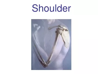
SHOULDER
SHOULDER. Kaan Yücel M.D., Ph.D. 27.December.2011 Tuesday. R egion of upper limb attachment to the trunk. P roximal segment of limb that overlaps parts of the trunk (thorax and back) and lower lateral neck. Shoulder includes P ectoral Scapular

SHOULDER
E N D
Presentation Transcript
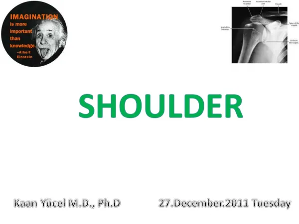
SHOULDER Kaan Yücel M.D., Ph.D 27.December.2011 Tuesday
Region of upper limb attachment to the trunk. Proximal segment of limb that overlaps parts of the trunk (thorax and back) and lower lateral neck. Shoulder includes Pectoral Scapular Deltoid regions of the upper limb lateral part (greater supraclavicular fossa) of lateral cervical region. Overlies half of the pectoral girdle.
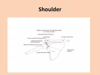
The pectoral (shoulder) girdle is a bony ring, incomplete posteriorly, formed by: Scapulae and clavicles completed anteriorly by the manubrium of the sternum (part of the axial skeleton).
Bone framework of the shoulder • Clavicle & scapula [form pectoral girdle-shoulder girdle-] • & • Proximal end of humerus
The superficial muscles of the shoulder trapezius & deltoid muscles together form the smooth muscular contour over the lateral part of the shoulder. Connect scapula to trunk & clavicle to arm. .
Joints The 3 joints in the shoulder complex : Sternoclavicular joint Acromioclavicular joint Glenohumeral joint
The two most superficial muscles of the shoulder are the trapezius and deltoid muscles. Together, they provide the characteristic contour of the shoulder: Trapezius attaches scapula & clavicle to the trunk Deltoidattaches scapula & clavicle to the humerus
Both the trapezius and deltoid are attached to opposing surfaces and margins of the spine of the scapula, acromion, and clavicle. The scapula, acromion, and clavicle can be palpated between the attachments of trapezius and deltoid.
SUPERFICIAL POSTERIOR AXIOAPPENDICULAR MUSCLES • EXTRINSIC SHOULDER MUSCLES • Trapezius & latissimus dorsi
Trapezius •Descending (superior) fibers elevate the scapula (e.g., when squaring the shoulders). •Middle fibers retract the scapula (i.e., pull it posteriorly). •Ascending (inferior) fibers depress the scapula and lower the shoulder. Movement to 180 degrees (elevation) is brought about by rotation of the scapula upwards by the trapezius and serratus anterior.
Latissimus dorsi • Passes from the trunk to the humerus and acts directly on the glenohumeral joint and indirectly on the pectoral girdle (scapulothoracic joint). • In combination with the pectoralis major, the latissimus dorsi is a powerful adductor of the humerus and plays a major role in downward rotation of the scapula in association with this movement.
DEEP POSTERIOR AXIOAPPENDICULAR MUSCLES EXTRINSIC SHOULDER MUSCLES Levator scapulae & rhomboids Provide direct attachment of appendicular skeleton to the axial skeleton
SCAPULOHUMERAL (INSTRINSIC SHOULDER) MUSCLES 6 scapulohumeral muscles Deltoid, teres major, supraspinatus, infraspinatus, subscapularis, and teres minor pass from scapula to humerus Act on the glenohumeral joint. All the intrinsic muscles but the deltoid and the subscapularis are muscles of the posterior scapular region.
Deltoid • Large and triangular in shape • Base attached to the scapula and clavicle • Apex attached to the humerus
Deltoid • Originates along a continuous • U-shaped line of attachment to clavicle & scapula • mirroring the adjacent insertion sites of the trapezius muscle. • Inserts into deltoid tuberosity on the lateral surface of the shaft of the humerus.
Deltoid Major function: Abduction of the arm beyond the initial 15° accomplished by supraspinatus
Deltoid • Innervation: Axillary nerve • branch of the posterior cord of the brachial plexus
Subscapularis • Thick, triangular muscle • Lies on the costal surface of the scapula • Originates from the subscapular fossa on the anterior surface of the scapula • Attaches to the lesser tubercle of the humerus
Subscapularis Primary medial rotator of the arm Adducts the arm. Joins the other rotator cuff muscles in holding the head of the humerus in the glenoid cavity during all movements of the glenohumeral joint (i.e., it helps stabilize this joint during movements of the elbow, wrist, and hand). Innervation: Superior and inferior subscapular nerves
POSTERIOR SCAPULAR REGION
The posterior scapular region occupies the posterior aspect of the scapula and is located deep to the trapezius and deltoid muscles. Contains 4 muscles pass between the scapula and proximal end of humerus: supraspinatus, infraspinatus, teres minor, teres major
The posterior scapular region also contains part of one additional muscle, the long head of the triceps brachii, which passes between the scapula and the proximal end of the forearm. This muscle, along with other muscles of the region and the humerus, participates in forming a number of spaces through which nerves and vessels enter and leave the region.
Supraspinatus & Infraspinatus • Originate from 2 large fossae, 1 above and 1 below the spine, on the posterior surface of the scapula. • Insert on greater tubercle of the humerus. • Supraspinatus initiates abduction of the arm. • Infraspinatuslaterally rotates the humerus.
Teres minor • A cord-like muscle • Originates from a flattened area of the scapula immediately adjacent to its lateral border below the infraglenoid tubercle. • Inserts on inferior facet of greater tubercle of humerus. • Laterally rotates the humerus and is a component of the rotator cuff.
Teres major • Originates from a large oval region on the posterior surface of the inferior angle of the scapula. • Attaches to the medial lip of the intertubercular sulcus on the anterior surface of the humerus. • Medially rotates and extends the humerus.
ROTATOR CUFF MUSCLES • 4 intrinsic shoulder muscles • Supraspinatus • Infraspinatus • Teres minor • Subscapularis (SITS muscles) Form a musculotendinous rotator cuff around the glenohumeral joint.
ROTATOR CUFF MUSCLES • Insertion exception: Supraspinatus Infraspinatus Teres minor greater tubercle Subscapularis lesser tubercle
ROTATOR CUFF MUSCLES Functional exception: All except supraspinatus are rotators of the humerus Supraspinatus, besides being part of the rotator cuff, initiates and assists the deltoid in the first 15° of abduction of the arm.
Gateways to the posterior scapular region Suprascapular foramen The route through which structures pass between the base of the neck and the posterior scapular region. Formed by suprascapular notch of scapula & superior transverse scapular (suprascapular) ligament, which converts the notch into a foramen.
Gateways to the posterior scapular region The suprascapular nerve passes through the suprascapular foramen; Suprascapular artery & suprascapular vein follow the same course as the nerve, but normally pass immediately superior to the superior transverse scapular ligament and not through the foramen.
Gateways to the posterior scapular region Quadrangular space Triangular space Triangular interval
Nerves The two major nerves of the posterior scapular region: Suprascapular & Axillary nerves originate from the brachial plexus in the axilla.
Suprascapular nerve Originates where? Base of the neck from superior trunk of brachial plexus Reach where through which space? Posterior scapular region through suprascapular foramen Which muscles innervates ? Supraspinatus muscle &infraspinatus
Axillary nerve Originates where? Posterior cord of brachial plexus Reach where through which space? From the posterior wall of axilla to posterior scapular region through quadrangular space Which muscles innervates ? Deltoid & teres minor
Axillary nerve Cutaneous branch? Superior lateral cutaneous nerve of the arm carries general sensation from the skin over the inferior part of the deltoid muscle.
Arteries • 3 major arteries in the posterior scapular region • Suprascapular artery • Posterior circumflex humeral artery • Circumflex scapular artery These arteries contribute to an interconnected vascular network around the scapula.
Formation of anastomosis around the surgical neck of humerus Posterior circumflex humeral artery anastomoses with anterior circumflex humeral artery and also with branches from: profunda brachii (brachial artery) suprascapular (subclavian artery) thoracoacromial (axillary artery) arteries .
Scapular anastomosis system • A system connecting each subclavian artery and the corresponding axillary artery, forming an anastomosis around the scapula. • It allows blood to flow past the joint regardless of the position of the arm. • It includes: • transverse cervical artery • (subclavian artery) • transverse scapular artery • (subclavian artery) • subscapular artery • (axillary artery) • branches of thoracic aorta . This collateral circulation allows for blood to continue circulating if the subclavian is obstructed.
Testing the deltoid muscle • To test the deltoid (or the function of the axillary nerve that supplies it), the arm is abducted, starting from approximately 15°, against resistance. • If acting normally, the deltoid can easily be seen and palpated.
Quadrilateral Space Syndrome Aclinical syndrome resulting from compression of the axillary nerve and posterior circumflex humeral artery in the quadrilateral space The passage of the axillary nerve backward from the axilla through the quadrangular space makes it particularly vulnerable here to downward displacement of the humeral head in shoulder dislocations or fractures of the surgical neck of the humerus.
Quadrilateral Space Syndrome Paralysis of the deltoid and teres minor Cutaneous branches of the axillary nerve, including the upper lateral cutaneous nerve of the arm a loss of skin sensation over the lower half of the deltoid muscle.
Movements of the shoulder girdle Abduction of the shoulder is initiated by the supraspinatus; the deltoid can then abduct to 90 degrees. Further movement to 180 degrees (elevation) is brought about by rotation of the scapula upwards by the trapezius and serratus anterior. As soon as abduction commences at the shoulder joint, so the rotation of the scapula begins.
Principal muscles acting on the shoulder joint Abductors Supraspinatus Deltoid Adductors Pectoralis major Lattisimus dorsi Extensors Teres major Lattisimus dorsi Deltoid (posterior fibres) Flexors Pectorali major Coracobrachialis Deltoid (anterior fibres) Medial rotators Pecroralis major Lattisimus dorsi Teres major Deltoid (anterior fibres) Subscapularis Lateral rotators Infraspinatus Teres minor Deltoid (posterior fibres)