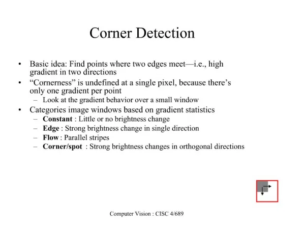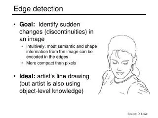
The Human Eye (part 2): Light detection
Students should be able to:<br>- describe the structure of the eye as seen in front view and in horizontal section

The Human Eye (part 2): Light detection
E N D
Presentation Transcript
Photoreceptors (retina) Connected to nerve- endings from optic nerve Nerve conveys impulses to brain when stimulated • • 2 kinds: 1. Rods 2. Cones
Question: • Have you experienced occasions when you are not able to see for a short period of time? • When does this situation normally take place?
Rods • Contain visual purple • Pigment concerned with vision in dim light • Light detection by visual purple by bleaching • From bright → dark place unable to distinguish objects for a while (because takes time for visual purple to reform) • Requires vitamin A (deficiency → night- blindness)
Cones Less sensitive to light Inefficient in dim light Concerned with bright light and colour vision • • • 3 types: (each contain a different pigment) 1. Red 2. Blue 3. Green • Pigments absorb light of different wavelengths • Taken together, cones allow us to see spectrum of colours
- yellow spot -where images are normally focused - highest conc. of cones - vision sharpest in bright light - No photoreceptors (not light sensitive) - blind spot formation
Fovea centralis • yellow spot • where images are normally focused • only contains cones • concentration of cones highest at this point • Therefore vision is sharpest in bright light when images are focused onto the yellow spot
Blind spot ▪ found immediately over optic nerve where it enters the eye ▪ has no photoreceptors (not sensitive to light) Blind spot
Formation of image on retina http://www.bausch.com/us/vision/teens/illusions/eyeworks.jsp
Image formed on retina 1. Inverted (upside down) 2. Reversed (back to front) 3. Diminished (smaller in size than the object being observed)
What are the similarities between the eye and a camera?
Processing by brain • Impulses produced by light falling on the rods and cones are transmitted via the optic nerves to rear of cerebrum • Brain interprets these impulses so we see the object the right way up and the right size