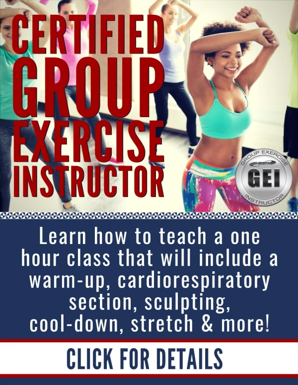
Group Exercise Instructor Certification Manual
Get the NESTA Group Fitness Teacher course manual. 100% Online Course u2013 Comprehensive Training u2013 Certification u2013 Complete Career System u2013 Ongoing Support. Become a group exercise leader and teach classes at your local gym, health club or fitness center. Help out at schools. You can also work with private clients in their homes. NESTA offers you many other courses, fitness trainer manual and career programs.<br>To learn more, please visit here: https://www.nestacertified.com/group-fitness-instructor/<br>

Group Exercise Instructor Certification Manual
E N D
Presentation Transcript
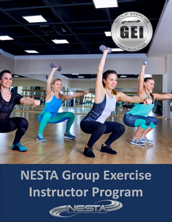
NESTA Group Exercise Instructor Program Instructor Program NESTA Group Exercise
Contents Section 1 Introduction Section 1.1 Introduction to Group Exercise . . . . . . . . . . . . . . . . . . . . . . . . . . . . . . . . . . . . . . . . . . . . . . . .1 Section 1.2 The Fitness Industry . . . . . . . . . . . . . . . . . . . . . . . . . . . . . . . . . . . . . . . . . . . . . . . . . . . . .5 Section 2 - The Science of Exercise Section 2.1 Kinesiology . . . . . . . . . . . . . . . . . . . . . . . . . . . . . . . . . . . . . . . . . . . . . . . . . . . . . . . . .7 Section 2.2 The Kinetic Chain . . . . . . . . . . . . . . . . . . . . . . . . . . . . . . . . . . . . . . . . . . . . . . . . . . . . . 19 Section 2.3 The Skeletal System . . . . . . . . . . . . . . . . . . . . . . . . . . . . . . . . . . . . . . . . . . . . . . . . . . . . 20 Section 2.4 The Muscular System . . . . . . . . . . . . . . . . . . . . . . . . . . . . . . . . . . . . . . . . . . . . . . . . . . . 27 Section 2.5 Functional Muscular Anatomy . . . . . . . . . . . . . . . . . . . . . . . . . . . . . . . . . . . . . . . . . . . . . . . 32 Section 2.6 Introduction to Exercise Physiology . . . . . . . . . . . . . . . . . . . . . . . . . . . . . . . . . . . . . . . . . . . . . 51 Section 2.7 Metabolism & Energy Systems . . . . . . . . . . . . . . . . . . . . . . . . . . . . . . . . . . . . . . . . . . 54 Section 2.8 The Cardiovascular System . . . . . . . . . . . . . . . . . . . . . . . . . . . . . . . . . . . . . . . . . . . . 61 Section 2.9 Proper Fuel: Nutrition Basics . . . . . . . . . . . . . . . . . . . . . . . . . . . . . . . . . . . . . . . . . . . . . . . . 66 Section 3 Group Exercise Instruction Section 3.1 Choreography & Class Building . . . . . . . . . . . . . . . . . . . . . . . . . . . . . . . . . . . . . . . . . . . . . . . 73 Section 3.2 Equipment and Tools for Teaching . . . . . . . . . . . . . . . . . . . . . . . . . . . . . . . . . . . . . . . . . . . . . . 87 Section 3.3 Music, Cueing & Corrections: . . . . . . . . . . . . . . . . . . . . . . . . . . . . . . . . . . . . . . . . . . . . . . . . 92 Section 3.4 Cueing & Learning Styles: . . . . . . . . . . . . . . . . . . . . . . . . . . . . . . . . . . . . . . . . . . . . . . . . . 97 Section 3.5 The “It Factor”. . . . . . . . . . . . . . . . . . . . . . . . . . . . . . . . . . . . . . . . . . . . . . . . . . . . . . 102 Section 4 Safety, Liability & Scope of Practice Section 4.1 Safety . . . . . . . . . . . . . . . . . . . . . . . . . . . . . . . . . . . . . . . . . . . . . . . . . . . . . . . . . 104 Section 4.2 Liability & Scope of Practice . . . . . . . . . . . . . . . . . . . . . . . . . . . . . . . . . . . . . . . . . . . . . . . 114 Resources. . . . . . . . . . . . . . . . . . . . . . . . . . . . . . . . . . . . . . . . . . . . . . . . . . . . . . . . 118 Image Credits . . . . . . . . . . . . . . . . . . . . . . . . . . . . . . . . . . . . . . . . . . . . . . . . . . . . . . 119
Section 1.1 Introduction to Group Exercise Section Objectives: Know the benefits of exercise for both the body and mind. impact classes with much less stress on the body. Hybrid classes combining hi-low moves became popular, and these classes still exist today. It seemed the perfect solu- tion. Students could get a great workout and avoid injury. Know the code of ethics for group exercise instructors. Welcome! Group Exercise began as “Aerobics” classes in the 1970’s when Kenneth Cooper coined the phrase “Aerobics.” In the beginning, classes consisted mainly of a warm- up, stretching, cardiovascular exercise with high impact moves, sculpting and a cool-down followed by a stretch. Many years later, Gin Miller brought a “step” to Reebok and everyone was “stepping.” These classes included the use of blocks or risers that would increase or decrease the height of the step to help accommodate various heights and fitness levels. These classes remained popular for a couple of decades, and you can still find a few on class schedules today. Some people, over time, began to experi- 1 GROUP EXERCISE INSTRUCTOR Eventually there were injuries; mainly of the feet, knees and back. The high impact classes transitioned into low
GROUP EXERCISE INSTRUCTOR Who is NESTA? NESTA is a professional fitness association offering a wide range of primary, advanced and specialized edu- cational courses and certification programs. NESTA was established in 1992 in Southern California. Today, NESTA is an international fitness association with over 55,000 members from around the world. Approximately 10% of our members are from outside the U.S. NESTA members and graduates have thriving careers in over 50 countries. Our certifications are accepted and used globally. We wel- come students from around the world. ence ankle and knee problems from traditional step class- es. Steps are found in most fitness facilities, however, but are used in classes other than step aerobics. They’re great props for Boot Camp, Circuit Classes and HIIT training. As Group Fitness classes evolved, along came: cycling, treadmill, kickboxing, circuit, aqua, BOSU, COREBOARD, functional fitness, Pilates, Yoga, Gliding, Core, Pole, Ta- bata, Kettlebell, and many more. Group Fitness evolved, and continues to evolve, every year. New inventions are made, old ones are brought back, and the desires of the people are being heard. 2 Who is this Program for? For anyone who wants to teach! Sure, it helps if you love fitness, people, helping and performing. But, if you have a love of teaching, this course is for you! One thing that never changes is the importance of the personality, skills and teaching techniques of the instruc- tor. If you ask someone why they rush from work to get to the club at a certain time for a class, they usually respond, “I love the instructor. She/he motivates me, encourages me to work hard and I really have a great time.” Mission Statement: This course will ready a future instructor to construct, and safely teach, one-hour group exercise classes that include: a warm-up, main workout based on title/theme, cool-down and stretch. Instructors will be able to “per- form” in each class to engage, encourage and excite par- ticipants to come back again and again. You could memorize this book from cover to cover, but it wouldn’t be enough to stand out above the rest. Only your unique style, care, commitment, passion and presentation can do that! So, let’s get started…
Goals of this Home Study: Our goal is to have you not only competent in group exer- cise instruction, but be a wildly successful member in the industry. The home study course is designed to be self- paced, easy to understand, concise and thorough. You’ll learn all about what is expected as an instructor, and you’ll have all the information needed to feel confident in interviews. You will need to read the manual, watch the videos and practice as you go. The anatomy portions will require some memorization. Each section will begin with “section objectives.” Think of these objectives as mini pre-tests along the way. If you are proficient in each objectives section, you stand a better chance of passing your final exam. Home Study Course Objectives: Students will develop a basic knowledge of anatomy, kinesiology and exercise physiology. 3 GROUP EXERCISE INSTRUCTOR Students will feel comfortable and confident in front of a class. Students will be able to develop and organize a fitness class. Students will be able to teach safe and effective classes using a variety of equipment. Students will be able to properly align bodies for each exercise. Students will develop leadership and teaching skills. Students will know basic functions of the muscles. Students will be able to understand the “Heart Rate Formula.” Students will be able to understand the “Perceived Exertion Chart.” Students will be able to identify the major muscle groups of the body and their functions as it applies to Group Fitness. Students will be able to identify major professional responsibilities of a group fitness instructor. Students will understand and identify the NESTA Code of Ethics. Students will become confident in cueing and teaching techniques. Students will be able to identify the musical beat and phrase used in teaching classes. Students will be able to teach at least 2 different exercises for each large muscle group. Students will be able to teach at least 1 stretch for each large muscle group. Students will make plans to take CPR training. Students will be able to identify special populations and seek professional advice as needed.
Code of Ethics for NESTA Certified Group Exercise Instructors: This code describes the appropriate conduct for all certified group exercise instructors working in the health, wellness and fitness industry. To provide exemplary instruction in a safe manner. To act with integrity, showing respect to all clients and fellow health, fitness and wellness profes- sionals. To always maintain professional boundaries, preserving the confidentiality of all students. To maintain a current certification through continuing education, and knowledge of the latest research and developments in the fitness industry. To conduct fitness, health and wellness business practices in accordance with all local, state and federal laws. To be current with all health, safety and first aid procedures. To be cognizant of professional limitations and seek the advice of other health professionals when appropriate. Benefits of a Group Fitness Class: GROUP EXERCISE INSTRUCTOR Physiological • Increases strength and endurance • Aid in weight loss and management • Improve focus and cognitive function • Minimize chronic disease • Improves cardiac efficiency • increased stroke volume • lowers resting heart rate • can increase HDL’s Psychological • Increases vigor • Decreases depression 4
Section 1.2 The Fitness Industry PASSION If you don’t have passion, nothing else will matter, so be sure that teaching and leading light you up! There’s no recipe for passion; you either have it or you don’t. You can overcome all the other “P’s” on this list, but without passion, nothing else really matters. If we are evolving, we are growing. Exercise trends come and go, and they certainly have in the fitness industry, but even trends spark something in people. Sometimes, a passing trend of mini trampolines or bouncing boots, might just inspire a couch potato to get up and try some- thing new. If the trend dies, at least they’ve been exposed to a gym setting, group classes and a friendly culture. It’s all about getting people from the couch to the gym. Once they’re there, we hope to win them over and make them lifers of the gym or whatever program comes out next. Good vs Great: PERSONALITY We all come to the table—or in this case, the stage—with our own unique personalities. It’s important to know yours! Are you an extrovert or an introvert? You can be a successful instructor, even as an introvert, so don’t let your self-realization scare you away from your passion. Just get to know your- self, and then remember to BE yourself. Copycats aren’t authentic; people see right through mimick- ing. If you’re bit shy, hone in on other traits that’ll make you shine. Maybe your empathy and warmth are your key strengths. Members will love that! If you’re loud and crazy, be aware of that energy too. Some people respond well to that, and others feel overwhelmed. It’s important to be yourself while considering things like: maintaining a good volume (too quiet or too loud are both problems), use of language (offensive language or jokes are just as problematic as shy commands that don’t convey authority). Are you there for the members and not yourself? People don’t care how much you know un- til they know how much you care! Showing off your knowledge of muscles or how many perfect burpees you can do won’t win people over. But if you notice a member who did 5 regular pushups after a week of modified, you’ve got a fan for life. It’s important to note that you won’t always be everyone’s cup of tea, no matter how much you try. Don’t get hung up on that! Variety is the spice of life, so don’t try to win everyone over or take it personally. If you are refining the best parts of yourself and focusing on your strengths in front of each class, your personality will get you far. 5 The deciding factor in your success in this industry rests on one question: Are you good, or are you great? Full classes, your name filling the schedule, happy members and a joyful experience rely on your greatness. The dif- ference between the two often comes down to: passion, personality, practice and perseverance. GROUP EXERCISE INSTRUCTOR If you don’t have passion, nothing else will matter, so be sure that teaching and leading light you up!
PRACTICE Do you know how Jerry Rice got the reputation as the hardest working football player in the NFL? Work ethic! He showed up before everyone, stayed later and ran his plays so that he could do them in his sleep. Much of what we do in the fitness industry is muscle memory, from the movements themselves, to choreography, to rhythmic timing. It can all be learned if you’re willing to practice. Do you have the discipline to outwork others? Nothing frustrates classes more than an unprepared teacher, or a teacher who doesn’t cue properly. There’s really no excuse for either. It simply comes down to practice. PERSEVERANCE Not unlike practice, perseverance is available to us all. How willing are you to stick with challenging choreography or memorizing a routine? How much time are you willing to invest in finding the perfect playlist for your class? Will you stick with your new career through bumps like low class attendance or constructive feedback? Great instructors welcome criticism to improve. Some even videotape them- selves to hear words they might overuse or to come up with more concise ways to cue. Every class is an opportunity to get better doing what you love, so stick to it, knowing that this is a marathon, not a sprint. GROUP EXERCISE INSTRUCTOR Think about it..... What do you think is your foremost personality feature which will help in developing your own group exercise program? Explain the pros and cons. 6 Which, if any, of the subjects in the NESTA Code of Ethics will be problematic for you? Why, and what do you need to do to resolve this? How do you display your passion? How do you plan to promote this or is it necessary to modify in any way?
Section 2 - The Science of Exercise In order to be an effective instructor, fitness professionals must have a strong understanding of the human body, how it moves, and how it adapts to exercise. This chapter will review the basics of kinesiology (the study of movement), functional anatomy and exercise physiology (how the body adapts to exercise). 7 GROUP EXERCISE INSTRUCTOR Section 2.1 Kinesiology Kinesiology is the scientific study of human movement. We start with a review of basic concepts in kinesiology, specifically basic movement terminology. It is vital that a fitness professional understand these terms and be capa- ble of properly applying them. The following terms pro- vide the foundation for properly describing human move- ment and the efficient application of exercise technique. This chapter also reviews functional anatomy. Traditional anatomy is based on the specific location of different bodily structures. Functional anatomy takes tradition- al anatomy a step further by considering muscle loca- tion as well as the movement produced by that muscle. Section Objectives: Be able to differentiate between anatomical position and anatomical neutral. Know all the planes of motion and be able to name an exercise done in each. Be able to describe a joint movement by the axis of rotation. Be able to successfully use anatomical directional terms. Be able properly define joint motion as based an anatomical movement descriptors.
Anatomical Position and Anatomical Neutral In order to describe the movement of the human body, a standardized reference point has been used for cen- turies known as Anatomical Position. Anatomical Position is a position of standing erect with the palms facing for- ward or externally rotated. There is another anatomical reference position with the palms facing the body known as Anatomical Neutral (a.k.a. Fundamental Starting Posi- tion). Kinesiology examines the way muscles interact. Therefore, it is essential for fitness professionals to study kinesiology to ensure that the exercises they prescribe to their students will help them achieve their goals and avoid injury. GROUP EXERCISE INSTRUCTOR 8
9 GROUP EXERCISE INSTRUCTOR Anatomical Position Anatomical Neutral
Planes of Motion We live in a three dimensional (3D) space. In order to describe human movement within three-dimensional space, three planes of motion are used. They are as follows: GROUP EXERCISE INSTRUCTOR 10 With knowledge of the three planes of movement, it is important to recognize that most human movement (outside of traditional weight lifting machines) is multi-planar and involves movement through more than one single plane. It would be insufficient to refer to the body in general terms (i.e., front, back, side, etc.) due to a need for clarity in reference points. A more efficient, scientific listing of basic anatomical movement descriptors is listed below.
The frontal plane divides the body into front and back halves (A.K.A. coronal plane) 11 GROUP EXERCISE INSTRUCTOR The sagittal plane divides the body into right and left halves The horizontal plane divides the body into top and bottom halves (A.K.A. transverse plane)
Anatomical Axis of Motion Each anatomical movement is achieved according to its joint makeup and is bound by the laws of motion around an axis. The Anatomical Axis of Motion describes a move- ment that occurs in a plane along an axis running perpen- dicular to the plane. Anatomical Directional Terms The front of the body relative to another reference point anterior (a.k.a. ventral) posterior (a.k.a. dorsal) The back of the body relative to another reference point The longitudinal axis contains movements that occur in the horizonal plane along the longitudinal axis. For exam- ple, the movement of turning one’s neck to look left and right is about the longitudinal axis. superior Above a reference point inferior Below a reference point A position relatively closer to the midline of the body A position relatively farther away from the midline A position closer to a reference point A position farther from the reference point medial The anterior-posterior axis includes movements in the frontal plane occur along the anterior-posterior axis. For example, the movement of raising one’s arm to the side as if completing a lateral raise (shoulder abduction) is about the anterior-posterior axis. lateral GROUP EXERCISE INSTRUCTOR proximal distal The coronal axis describes movements that occur in the sagittal plane occur along the coronal axis. For example, the movement kicking a leg forward (hip flexion) is about the coronal axis. unilateral Refers to only one side superficial Near the surface 12 deep Further beneath the surface cephalic Toward the head caudal Toward the bottom supine Lying on one’s backside prone Lying face down Anatomical movement descriptors, anatomical position and axes of motion are all important facts to know when becoming a fitness professional. These standards have been formulated to allow communication between professionals. Knowing this terminology will help you communicate with personal trainers, physicians, athletic trainers, physical therapists, chiropractors and other fitness professionals to ultimately make you a better and more knowledgeable group exercise instructor.
Think about it..... Can you name the dominant plane of motion in each of the images below? 13 GROUP EXERCISE INSTRUCTOR Frontal; Frontal; Sagittal; Frontal; Frontal; Sagittal; Frontal; Frotal and Sagittal; Sagittal; Sagittal; Sagittal; Sagittal; Sagittal; Sagittal; Sagittal; Sagittal and Horizontal From the top right....
sagittal plane movements (coronal axis) A bending movement where the relative angle between two adjacent segments decreases flexion flexion GROUP EXERCISE INSTRUCTOR flexion A bending movement where the relative angle between two adjacent segments increases extension flexion extension extension 14 flexion extension extension extension Hyperextension is a movement which continues past anatomical position. (a.k.a. Extension beyond neutral.) flexion
frontal plane movements (anterior-posterior axis) Movement away from the midline of the body abduction Movement towards the midline of the body adduction abduction abduction 15 GROUP EXERCISE INSTRUCTOR adduction adduction transverse plane movements (longitudinal axis) The rotation of a body part towards the midline (in- side) of the body The rotation of a body part away from the midline of the body internal/ medial rotation external rotation external/ lateral rotation internal rotation
Specialized Movement Descriptors positions affecting joint ROM and Movements of the Scapula stability protraction Abduction of the scapula GROUP EXERCISE INSTRUCTOR retraction Adduction of the scapula 16 elevation Raising of the scapula (shrug) depression Lowering of the scapula
Other specialized movements: lateral flexion (also known as lateral blending) spinal movement in the frontal plane A cone-shaped movement by the body. Circumduction does not require internal and external rotation. circumduction dorsiflexion dorsiflexion and plantar flexion 17 Only occurs in the foot GROUP EXERCISE INSTRUCTOR plantarflexion At wrist: Movement of the fore- arm where the palm rotates to face forward from neutral to ana- tomical position at the radioulnar joint pronation supination supination At foot-ankle: Supination is a tri- plane motion which combines the movements of inversion, ad- duction, plantarflexion At wrist: Movement of the fore- arm where the palm rotates to face backward When trying to remember the motion of supination in the hand, there are a few tricks to help you remember the posi- tion. Since the palm is up in a cupped position, you can think of supination as holding a cup of “soup.” Also, the word “up” is in supination, so your palm is up. pronation At foot-ankle: Pronation is a tri- plane motion consisting of simul- taneous movements of eversion, abduction, dorsiflexion
Think about it..... Can you name the movement descriptors for each joint in the image below? GROUP EXERCISE INSTRUCTOR 18 shouders are abducted. Cervical rotation to the left Slight plantar flexion in both ankles. Left knee is flexed. Left hip is flexed, abducted and externally rotated. Right hip is abducted and externally rotated. Both From the ground up....
Section 2.2 The Kinetic Chain Section Objectives: Be able to define the kinetic chain and its importance to human movement. Be albe to list the three bodily systems that make up the kinetic chain. Be able to describe the integrative nature of the kinetic chain. What is the Kinetic Chain? The study of exercise physiology must begin with an understanding of how the body moves. To fully under- stand human movement, one must first understand the structure and function of bodily muscle and its interde- pendence upon the nervous and skeletal systems. Much like the checks and balances that exist in human organi- zation to ensure efficient operation, there is a delicate balance within the system known as the Kinetic Chain. The kinetic chain is composed of the nervous system, muscular system and skeletal system. The systems of the Kinetic Chain must function efficiently for proper func- tioning of the entire body. Kinetic is usually defined as relating to, or produced by motion. Therefore, the kinetic chain is the “chain” of systems that “link” together to create human movement. 19 GROUP EXERCISE INSTRUCTOR Courtesy of 3D Science Fitness professionals realize that training involves more than just muscles. All systems of the body must work together as an integrated unit to perform even the most basic tasks. While we define the kinetic chain as three separate sys- tems which work together to create movement, the en- tire body must work as an integrated unit. For example, the respiratory and digestive systems must import the fuel sources, which power physiological movement. The cardiovascular system must transport fuel to the muscles before movement can occur.
Section 2.3 The Skeletal System Section Objectives: Be able to describe the various roles of the skeletal system. Be able to explain the materials that make up bone and the structural organization that allows bone to withstand loads. Be able to explain how bone adapts to load. GROUP EXERCISE INSTRUCTOR Be able to categorize joints based on structure and movement capabilities. Be able to describe joint positions that affect range of motion and stability. 20 Be able to describe the components that affect joint stabilization during exercise. Introduction and Goal The human skeleton consists of 206 bones. We are ac- tually born with more bones (about 300), but many fuse together as we grow during childhood. These bones support our body and allow us to move. Bones store cal- cium, a mineral that helps bones stay strong. Calicum is needed for proper funtioning for both nerves and muscles as well. Bones manufacture blood cells and store import- ant minerals for the body. Males and females have slightly different skele- tons, as well as having a different elbow angles. Males have slightly thicker and longer legs and arms to accomodate their “hunter-gathering” role; females have a wider pelvis and a larger space within the pelvis to make the whole birthing process possible! The Goal of this section is to give Group Exercise In- structors an understanding how the skeletal system is designed for both support and movement. In addition, you will be able to educate your athletes on basic joint structure and function, which is essential for safety and efficiency in group exercise classes. The longest bone in our body is the femur (thigh bone). The smallest bone is the stirrup bone located inside the ear. Each hand has 26 bones in it. The nose and ears are not made of bone; they are made of cartilage, which is a flexible substance that is not as hard as bone.
roles of the skeletal system levers For motion and restriction of motion The skeletal system is passive and can only move by muscular ac- tion. Active systems (such as the muscular system) can only move with the support of the skeletal system. The simplest example is the ribcage which provides protection for internal organs such as the lungs and heart. The spongy section of bone is composed of calcium, phosphate and magnesium support protection storage blood cell formation In the bone marrow structure and function of bone 21 GROUP EXERCISE INSTRUCTOR short The bone is wider than it is long; ex: tarsals and carpals flat ex: pelvis irregular ex: vertebrae Cylindrical shaft composed mostly of compact bone surrounding a cavity of bone marrow Dilated ends, composed of spongy bone surrounded by thin layer of compact bone Disk of cartilaginous cells separating diaphysis and epiphysis - site of longitudinal growth Fibrous sheath, surrounds long bone, site of attachment to muscle tissue diaphysis epiphysis long The bone is longer epiphyseal plate than it is wide (bones periosteum of the appendages); ex: femur, humerus sesamoid ex: patella
bone tissue • One of the body’s hardest structures • 60-70% of bone are made of the minerals calcium and phosphate, and the connective tissue collagen • Collagen gives bone it’s ability to withstand tensile (pulling) loads as well as bone’s ductile prop- erties • Minerals give bone brittleness and the ability to withstand compressive loads • Composed of 25-35% water GROUP EXERCISE INSTRUCTOR 22 architecture of bone compact/ cortical bone Dense, compact tissue on the exterior of bone that provides strength and stiff- ness to the skeletal system. Lattice-like and having high porosity, it is capable of high energy storage. Found on interior of bones and in the ends of long bones. Made up of small flat pieces of bone (trabeculae) that adapt to the direction of the imposed stress/force on bone. spongy/ cancellous bone
Wolff’s Law Wolff’s Law states that bone will adapt and grow in line with resistance forces. Bones respond best to com- pressive forces. Lateral forces may cause the greatest stress and potential damage. However, the bone will still have adaptive abilities. Such forces may have the great- est risk in young athletes/individuals with epiphyseal plate (growth plate) slippage during the years of greatest pre-pubescent growth between the ages of 11 and 14. Epiphyseal plate slippage may inhibit the growth of bone at the affected joint but will not necessarily affect growth of the entire body. Resorption and Deposit of Bone Bone is highly adaptive, self-repairing and can alter its properties and configuration in response to mechanical demand. Wolff’s Law states: “Every change in the form and function of a bone or of their function alone is followed by certain definitive changes in their internal architecture, and equally definite secondary alteration in their external conformation, in accordance with mathematical laws.” In other words, bone adapts to the mechanical stress placed upon it. Large volumes of bone are removed through bone resorption and replaced through deposit. In young adults, the bone deposits equal the bone resorption, and total bone mass is fairly constant. Weight training will increase the amount of bone deposits. Osteoporosis occurs when bone resorption exceeds bone deposits. 23 GROUP EXERCISE INSTRUCTOR Bones require mechanical stress that is created by physical activity in order to grow and become strong. This stress must be performed on a daily basis. Bone loss following a decrease in activity may be significant without sufficient stress.
Structure and Function of Joints While muscular injuries are fairly common, joint injuries are often the most devastating, causing long-term problems. Unlike most muscular inju- ries, joint injuries are more likely to last a lifetime. Hence, it is imperative that fitness professionals understand joint structure and function to prevent the occurence of such devastating injuries. A Joint is an articulation between two bones, which is used to connect one component of a structure with one or more other components. The design of a joint is determined by its function and the nature of its com- ponents. Once a joint is constructed, the structure of the joint will determine its function. Joints that serve a single function are less complex than joints that serve multiple functions. Joint typing is based on the type of materials and the methods used to unite bony compo- nents. GROUP EXERCISE INSTRUCTOR joint types synarthrodial immovable joints (pel- vis) amphiarthroses 24 semi-movable joints Courtesy of 3DScience.com (sacroiliac (SI) & pubis) 5 characteristics of synovial (diarthrodial) joints 1. Articulating bones are separate (not fused) 2. Held together by a capsule of fibrous connective tissue 3. Joint capsule is lined by a synovial membrane 4. Articulating surfaces are covered by cartilage (usually hyaline), but can be fibrocartilage such as the sternoclavicular joint or temporomandibular joint (atypical of synovial joints) 5. A small amount of synovial fluid can be found between articulating bones. Synovial diarthrodial freely movable joints (hip and knee) fluid is a liquid that reduces friction in the joint. The synovial fluid changes viscosity (fluid friction) in response to joint movement (this is one of the many reasons for an efficient warm-up prior to exercise).
movement diarthrodial joint type example (Degrees of Freedom are the number of independent movements allowed at a joint) Two saddle shaped surfaces allowing two degrees of freedom saddle carpometacarpal joint of the thumb Biaxial, one plane of movement that domi- nates two planes of motion 25 condyloid GROUP EXERCISE INSTRUCTOR tibiofemoral joint, Inter- phalangeal joint hinge One degree of freedom humeroulnar Flat surface that allows translation between two bones plane “gliding” acromioclavicular joint pivot One degree of freedom atlantoaxial joint “Ball and Socket;” three degrees of freedom, freely movable, allowing motion in all three planes of human movement multiaxial hip joint
positions affecting joint ROM and stability A joint position in which there is maximal contact between two joint surfac- es. The majority of ligaments are taut, forcing two bones to act as a single unit. For example, a “locked” (fully extended) knee or elbow is very stable, but is vulnerable to injury due to decreased mobility at either end range of muscular length (excessively short or long). A joint position with less than maximal contact between joint surfaces and in which contact areas are frequently changing. For the most part, any other joint position that is not close-packed (not locked). Less stable than closed-packed, but not as susceptible to injury due to increased mobility and muscular ability. characteristics of ligaments close-packed loose-packed GROUP EXERCISE INSTRUCTOR • Connect bone to bone • Comprised of collagen which is arranged to handle tensile and shear loads • Maintains contact surfaces at the joint by either limiting or completely preventing “unwanted” (potentially harmful) motion • Passive stabilizers of the joint which are not meant to be stretched!!! 26 forms of joint stabilization during exercise Internal passive stabilization • Performed by noncontractile components (ex: Ligaments) • Needed when there is no anatomical muscular support (ex: lateral knee), • Physiologically muscular support (i.e. no motor pattern), or during ac- tive insufficiency passive External passive stabilization • Performed by an outside force (ex: Bench) Static • Achieved by an isometric contraction of various muscular groups to pre- vent movement at a joint • While not very functional, it is often required for proper force distribu- tion Dynamic • This is stabilization that takes place throughout a range of motion. The prevention of undesirable motions is controlled by dynamic stabilizers in order to maintain relative positions of the appropriate segments. active (generated from muscular systems)
Section 2.4 The Muscular System Structure and Function of Muscular System A muscle is a structure composed of tissues which produce the movements of the body through mus- cular contractions. Muscles that are capable of volun- tary contraction are referred to as striated or skeletal muscle, while involuntary muscles, except the heart, are termed smooth muscle. Section Objectives: Be able to describe the three diffenent types of muscle tissue. Be able to describe how muscles are stimulated by the nervous system. Skeletal Muscle Skeletal muscle or striated muscle cells are some of the largest cells in the body and it is their job to cre- ate movement. They do so by contracting (shortening), which pulls the opposite ends of the muscle together. There are over 600 muscles in the human body, and Be able to explain the relationships of muscle fiber type to muscle function. Introduction and Goal There are three different types of muscle tissue: 27 more than 400 are skel- etal muscles. Fascia is a sheet or band of fibrous connective tissue which separates and contains muscles within com- partments. A tendon is a fibrous cord (consist- ing primarily of collagen) which attaches muscle to bone, although fascia can also act as a site for muscle attachment. GROUP EXERCISE INSTRUCTOR muscle tissue Involuntary actions (auto- nomic nervous system). No striations. Voluntary movement (somatic nervous system). Has striations. An involuntary muscle but has some striations. Digestive tract, bladder, uterus, blood vessel walls smooth All muscles controlling movement skeletal cardiac Heart only The skeletal muscular system is the anatomical system most affected by exercise. The framework for the body is provided by the bones and joints. However, movement is enabled by the contraction and relaxation of specific muscles. The goal of this section is to give the student a fundamental understanding of how muscles are able to contract and how they adapt to the stimulus of exercise. In addition to the fascia, there are three layers of con- nective tissue which surround, protect, and enable sep- arate innervation of muscle fibers (muscle cells) within a muscle. The outermost layer is the epimysium, which completely surrounds muscle. The next layer groups Why do we care? By understanding how muscle, bone and connective tissue are interdependent to one another, we will not only understand the rationale in creating safer programs but also the underlying causes of injuries such as tendonitis (inflammation of the tendon) and plantar fasciitis (inflammation of the fascia at the arch of the foot).
Sarcomere and the Sliding Filament Theory The sarcomere is composed of thin filaments (chains of globular actin) and thick filaments (organized bundles of myosin). During muscular contractions, these fila- ments slide over each other with the resulting action be- ing that the sarcomeres shorten together, thereby caus- ing the muscle to contract as a whole. Skeletal muscle is composed of bundles of long, parallel fibers (cells). Embedded in the muscle fibers are fila- ments called myofibrils which further divided into sar- comeres. Sarcomeres are the smallest contractile unit of skeletal muscle. Muscle fibers are called striated because they have striations of light and dark bands created by the repeating actin and myosin filaments in the sarcomeres. Muscles are divided by layers of connective tissue to en- able it to be innervated from smaller to larger numbers of fibers and sizes. A motor neuron and all the muscle fibers it activates is known as a motor unit. According to the size principle of recruitment, smaller numbers of fibers will be selected before larger numbers of fibers to pre- serve muscular energy by firing smaller motor neurons, followed by larger motor neurons. Furthermore, once a muscle fiber is innervated, it is either on or off, there can be no partial contraction; this is the all-or-none theory of muscle fiber activation. GROUP EXERCISE INSTRUCTOR Action Potentials Muscle fibers contract in response to an electrical signal sent by the motor neuron. This signal is known as an action potential. The signal is sent down the motor neu- ron to its terminal ending on a muscle fiber known as the motor end plate. Where the motor end plate meets the muscle fiber is the neuromuscular junction. 28 Courtesy of 3DScience.com If the signal is strong enough, an action poten- tial of the muscle fiber is generated. This causes the fiber to contract.
structure of the sarcomere Defines boundary of single sarco- mere Runs down center of sarcomere Z-lines M-line I-band H-zone Contains thin filaments only Contains thick filaments only Contains thick filaments in their entirety without any overlapping portions of thin filaments A-band 29 GROUP EXERCISE INSTRUCTOR roles of skeletal muscle prime mover or agonist A muscle that acts directly to bring about a desired movement through concentric muscle action assistant mover or synergist A muscle that assists indirectly to bring about a desired movement A muscle responsible for opposing the concentric muscle action of the agonist (more susceptible to injury). antagonist stabilizer A muscle responsible for stabilizing an adjacent segment
GROUP EXERCISE INSTRUCTOR muscle fiber types 30 RED, slow twitch, aerobic fibers These fibers are fatigue-resistant. They are rich in capillaries and myoglobin (enhancing oxygen delivery). They also have increased number of mitochondria which enhances their ability to oxidize fats. Muscles containing a predominant amount of slow twitch muscle fibers tend toward stabilizing functions rather than prime movers of muscle tissue. Individuals with a greater number of Type I fibers tend to excel in endurance related activities that last longer than 3 minutes. type I fibers PINK, moderately fast twitch These fibers have traits of both Type I and Type IIB. Individuals with a greater number or more conditioned Type IIA fibers tend to excel in activities lasting between 30 seconds and 2 minutes with a moderate level of strength and aerobic capacity capabilities. type IIA fibers WHITE, fast twitch, anaerobic fibers These fibers are larger in diameter and well suited for brief, powerful contractions. They have little mitochondria and small capillary beds. These fibers tend to be prime movers of joints. Individuals with a greater number of Type IIB fibers tend to excel in highly explosive activities such as maximal lifts and sprinting or jumping. These are also the fibers that have the greatest potential for hypertrophy. NOTE: Historically these fibers are known as “type IIB” in humans. However, based on newer techniques to classify muscle fibers based on the type of myosin they have, the newer designation of “type IIx” is now used. type IIB fibers a.k.a. “type IIx”
Fried Chicken If you want to get a clear idea of the difference between muscle fiber types, look no farther than your nearest bucket of fried chicken. Chickens tend to either sit or walk all day, so their legs and thighs need endurance (the dark meat are Type I fibers). On the other hand, to avoid predators, chickens can explosively use their breast and wing muscles (the white meat - Type IIB fibers) to get away (that is the extent of their flying capabilities). net muscle actions Definition: Shortening of a muscle fiber against a load • “Raises” the load • “Accelerates” the load • Ex: Upward phase of a dumbbell bicep curl Definition: Lengthening of a muscle fiber against a load • “Lowers” the load • “Decelerates” the load • Ex: Downward phase of a dumbbell bicep curl Definition: Where a muscle neither lengthens nor shortens against a load (no net joint movement). A co-contrac- tion of agonist and antagonist muscles. • “Stabilizes” the load • Ex: Holding the elbow still at 90˚ in a dumbbell bicep curl concentric Training Adaptations to the Muscular System The increase in the size of muscle is known as hyper- trophy. Hypertrophy is an increase in the size of my- ofibrils inside muscle fibers. Hyperplasia is an increase in the number of muscle cells in the body with a cor- responding increase in muscular size. The phenomenon of hyperplasia is extremely rare, and has only been seen in a few highly advanced bodybuilders and professional athletes. Muscle atrophy on the other hand, refers to the wasting or loss of muscle tissue resulting from disease or lack of use. eccentric 31 GROUP EXERCISE INSTRUCTOR isometric events takes place during all movements, especially during the initial stages of training (motor learning/motor ability stage). Muscular Strength and Endurance There are many types of strength that an individu- al may need in order to improve performance and/ or function. In order to understand the benefits of ex- ercise, we must first define and describe the different types of strength. Is it easier to accelerate, stabilize or decelerate a load? While running, is it easier to speed up or slow down? It is easiest to speed up and the most difficulty is found in attempting to slow down. It is also much harder to stabilize a load in space than it is to create movement with a load in most situations. Therefore, eccentric is referred to as the “strongest” muscular action and concentric the “weakest” of the three. Keep in mind that most injuries occur eccentrically as the muscles attempt to control the downward phase or to deceler- ate load. When running, jumping or lifting, injury often occurs because of an inability to decelerate due to the build-up of forces from the previous acceleration and the acceleration of gravitational or other resistance forces. Neural Factors Influencing Strength Gains Exercise elicits a training stimulus: One effect is struc- tural (hypertrophy of muscles and connective tissues); the other is the functional aspect either within a muscle (intramuscular coordination) or within a group of mus- cles (intermuscular coordination). During any movement, certain muscles will be activated as a primary, secondary, and/or stabilizers to coincide with facilitation (actual recruitment) or inhibition (shut off). This sequence of
Section 2.5 Functional Muscular Anatomy Section Objectives: Know the major muscles and their actions in the following regions: Muscles of the Lower Leg Muscles of the Thigh (Quadriceps) Muscles of the Thigh (Hamstrings) Muscles of the Hip (Gluteal Muscles) Muscles of the Hip (Hip Flexors) GROUP EXERCISE INSTRUCTOR Muscles of the Hip (Adductor Group) Muscles of the Hip (Hip Abductors) Muscles of the Vertebral Column (Deep to Superficial) Muscles of the Vertebral Column (Posterior Muscles) 32 Upper Extremity Musculature Muscles of the Shoulder Girdle Muscles of the Shoulder Girdle (Rotator Cuff) Muscles of the Upper Arm All About Muscles There are three general types of muscular tissue. These types can be designated by location, appearance, and control. There are three main parts to a muscle: Origin (or head) The less moveable attachment NOTE: there can be more than one origin Belly The bulging part of a muscle Insertion The moveable attachment
33 GROUP EXERCISE INSTRUCTOR
Below is a list of the major muscles of the human body which are the primary concern of a group exercise instructor. REMEMBER, this is not an all-inclusive list and every fitness professional should have a high-quality anatomy book in their library. In addition, the primary functions listed are the actions of the muscles when they contract concentrically (shorten under load). REMEMBER, muscles do not just work concentrically to “lift a load!!” Muscles can also act eccentrically to decelerate the opposite motion as well as statically (isometrically) in order to stabilize the joints that they cross over. In other words, when you read the primary functions of each muscle – remember that the muscle will also fire to “lower the load” (the opposite motion) and stabilize motion (or lack of motion).** major movers of the lower leg and their primary funcitons 35 soleus plantarflexion GROUP EXERCISE INSTRUCTOR gastrocnemius plantarflexion and knee flexion peroneus (fibularis) plantarflexion and eversion longus anterior tibialis dorsiflexion and inversion
Think about it..... From the previous list, which muscles would be contracting during a calf raise? GROUP EXERCISE INSTRUCTOR 36 The movement is primarily performed by the gastrocnemius and soleus, with some assistance from the peroneus longus as an assistant mover.
thigh muscles (quadriceps) rectus femoris knee extension, hip flexion 37 vastus intermedius GROUP EXERCISE INSTRUCTOR (deep to rectus knee extension femoris) vastus lateralis knee extension knee extension and medial glide vastus medialis of patella
thigh muscles (hamstrings) biceps knee flexion, hip extension femoris and external rotation GROUP EXERCISE INSTRUCTOR knee flexion, hip extension semitendinosis and internal rotation 38 knee flexion, hip extension semimembranosus and internal rotation Although muscle groups such as the quadriceps and hamstrings work together to complete the same movement, they also have different actions, independent of each other. While the vastus medialis primarily works to extend the knee, it also has an important role in stabilizing the patella on the medial side, where it attaches. This is why understanding both the origins and insertions of muscles are important, in addition to their actions.
hip muscles (gluteal) external rotation, hip extension gluteus maximus upper-half - hip abduction lower-half - hip adduction 39 GROUP EXERCISE INSTRUCTOR abduction, assists in hip flexion gluteus medius anterior fibers - internal rotation posterior fibers - external rotation abduction, internal rotation and gluteus minimus assists in hip flexion
hip muscles (hip flexors) hip flexion, lumbar flexion, psoas major external rotation and slight hip abduction GROUP EXERCISE INSTRUCTOR 40 hip flexion, lumbar flexion and Iliacus external rotation When combined, the Iliacus and Psoas Major are also known as the Iliopsoas.
hip muscles (adductors) adductor brevis adduction and hip flexion 41 GROUP EXERCISE INSTRUCTOR adductor longus adduction and hip flexion adduction (some fibers contribute to both internal adductor magnus and external rotation as well as hip flexion and extension)
other hip muscles hip adduction, internal gracilis rotation and knee flexion GROUP EXERCISE INSTRUCTOR hip flexion, abduction, tensor fascia latae internal rotation 42 hip flexion, abduction, external rotation. knee sartorius flexion and internal rotation Fun Fact: The sartorius is the longest muscle in the human body. It is also known as the “tailor’s muscle,” for the crossed leg position that a tailor often sits in.
trunk musculature anterior vertebral column muscles forced expiration and in- creased intra-abdominal pressure (stiffens the trunk to prevent the spine from buck- ling under compressive loads) transverse transverse abdominus abdominus rectus abdominus Flexion, posteriorly tilts pelvis and increases intra-abdominal pressure. Although the rectus abdominus is responsible for flexing the “spine,” it does not actually attach to the vertebrae. The rectus abdominus attaches to the pubis and sternum. rectus 43 abdominis GROUP EXERCISE INSTRUCTOR flexion, ipsilateral (same side) spinal rotation and increased intra-abdominal pressure internal oblique external oblique Fiber direction - run down and out flexion, contralateral (opposite side) spinal rotation, tilts pelvis posteriorly and increased internal oblique external intra-abdominal pressure. oblique Fiber direction - think of hands in pockets (i.e. down and in) quadratus lateral flexion of the vertebral column and laterally tilts pelvis lumborum
posterior vertebral column muscles - erector spinae muscles iliocostalis cervicis longissimus capitis most lateral iliocostalis thoracis longissimus cervicis extend the vertebral column and bend it laterally. iliocostalis group (cervicis, thoracis, lumborum) GROUP EXERCISE INSTRUCTOR longissimus thoracis longissimus group (capitis, cervicis, thoracis) extend the vertebral column and head, rotate head. iliocostalis lumborum 44 spinalis capitis spinalis cervicis most medial extend the vertebral column. spinalis group (capitis,cervicis, thoracis) spinalis thoracis
upper extremity musculature shoulder girdle muscles (i.e. muscles that move the scapula - shoulder blade) Trapezius upper fibers scapular elevation middle fibers scapular retraction scapular depression (upward rotation occurs in conjunction with serratus anterior) 45 lower fibers GROUP EXERCISE INSTRUCTOR rhomboid scapular retraction (adduction) and some elevation major and minor downward or medial rotation of the scapula and scapular elevation levator scapulae serratus protraction and upward rotation of the scapula anterior
shoulder girdle muscles (continued) stabilizes and depresses subclavius the shoulder girdle GROUP EXERCISE INSTRUCTOR depresses the scapula and pectoralis minor pulls it anteriorly 46 Think about it..... Of the muscles mentioned, which would be be the prime movers during a shrug? The prime mover of the shrug is the upper fibers of the trapezius.
deep muscles of the shoulder joint - the rotator cuff these muscles work together to maintain integrity and alignment of the glenohumeral joint Stability of the shoulder joint during swimming and throwing movements. Some abduction/external rotation. supraspinatus 47 External rotation of the humerus. In extension, helps move the humerus posteriorly. GROUP EXERCISE INSTRUCTOR infraspinatus External rotation of the humerus. In extension, it helps move the humerus posteriorly. teres minor Internal rotation of the humerus. In extension it helps move the humerus posteriorly. In adduction, it helps move the arm toward the body. subscapularis