Download

1 / 123
1.28k likes | 2.22k Vues
NUCLEAR MAGNETIC RESONANCE. Nuclear magnetic resonance (NMR) is the spectroscopic study of the magnetic properties of the nucleus of the atom. The protons and neurons of the nucleus have a magnetic field associated with their nuclear spin and charge distribution. .

E N D
NUCLEAR MAGNETIC RESONANCE
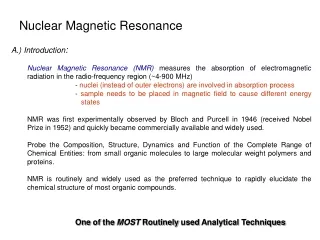
Nuclear magnetic resonance (NMR) is the spectroscopic study of the magnetic properties of the nucleus of the atom. • The protons and neurons of the nucleus have a magnetic field associated with their nuclear spin and charge distribution.
Resonance is an energy coupling that causes the individual nuclei, when placed in a strong external magnetic field, to selectively absorb, and later release, energy unique to those nuclei and their surrounding environment. • The detection and analysis of the NMR signal has been extensively studied since the 1940s as an analytic tool in chemistry and biochemistry research. • NMR is not an imaging technique but rather a method to provide spectroscopic data concerning a sample placed in the device.
In the early 1970s, it was realized that magnetic field gradients could be used to localize the NMR signal and to generate images that display magnetic properties of the proton, reflecting clinically relevant information. • As clinical imaging applications increased in the mid-1980s, the “nuclear” connotation was dropped, and magnetic resonance imaging (MRI), with a plethora of associated acronyms, became commonly accepted in the medical community.
MRI is a rapidly changing and growing image modality. • The high contrast sensitivity to soft tissue differences and the inherent safety to the patient resulting from the use of non ionizing radiation have been key reasons why MRI has supplanted many CT and projection radiography methods. • With continuous improvements in image quality, acquisition methods, and equipment design, MRI is the modality of choice to examine anatomic and physiologic properties of the patient
There are drawbacks, including • High equipment and siting cost, • Scan acquisition complexity, • Relatively long imaging times, • Significant image artifacts, and • Patient claustrophobic problems.
Magnetism • Magnetism is a fundamental property of matter; it is generated by moving charges, usually electrons. • Magnetic properties of materials result from the organization and motion of the electrons in either a random or a nonrandom alignment of magnetic “domains,” which are the smallest entities of magnetism.
Atoms and molecules have electron orbitals that can be paired (an even number of electrons cancels the magnetic field) or unpaired (the magnetic field is present). • Most materials do not exhibit overt magnetic properties, but one notable exception is the permanent magnet, in which the individual domains are aligned in one direction.
Magnetic susceptibility describes the extent to which a material becomes magnetized when placed in a magnetic field. • The induced internal magnetization can oppose the external magnetic field and lower the local magnetic field surrounding the material. • On the other hand, the internal magnetization can form in the same direction as the applied magnetic field and increase the local magnetic field.
Three categories of susceptibility are defined: • Diamagnetic, • Paramagnetic, and • Ferromagnetic.
Diamagnetic materials have slightly negative susceptibility and oppose the applied magnetic field. • Examples of diamagnetic materials are calcium, water, and most organic materials (chiefly’ owing to the diamagnetic characteristics of carbon and hydrogen).
Paramagnetic materials have slightly positive susceptibility and enhance the local magnetic field, but they have no measurable self-magnetism. • Examples of paramagnetic materials are molecular oxygen (O2), some blood degradation products, and gadolinium-based contrast agents.
Ferromagnetic materials are “superparamagnetic”—that is, they augment the external magnetic field substantially. • These materials can exhibit “self-magnetism” in many cases. • Examples are iron, cobalt, and nickel.
Unlike the monopole electric charges from which they are derived, magnetic fields exist as dipoles, where the north pole is the origin of the magnetic field lines and the south pole is the return. • One pole cannot exist without the other.
As with electric charges, “like” magnetic poles repel and “opposite” poles attract. • The magnetic field strength, b, (also called the magnetic flux density) can be conceptualized as the number of magnetic lines of force per unit area.
The magnetic field drops off with the square of the distance. • The SI unit for b is the tesla (T), and as a benchmark, the earth’s magnetic field is about 1/20,000 T. • An alternate unit is the gauss (G), where 1 T = 10,000 G.
Magnetic fields can be induced by a moving charge in a wire.
The direction of the magnetic field depends on the sign and the direction of the charge in the wire, as described by the “right hand rule”: • The fingers point in the direction of the magnetic field when the thumb points in the direction of a moving positive charge (i.e., opposite the direction of electron movement). • Wrapping the current-carrying wire many times in a coil causes a superimposition of the magnetic fields, augmenting the overall strength of the magnetic field inside the coil, with a rapid falloff of field strength outside the coil.
Direct current (DC) amplitude in the coil determines the overall magnitude of the magnetic field strength. • In essence, this is the basic design of the “air core” magnets used for diagnostic imaging, which have magnetic field strengths ranging from 0.3 to 2.0 T, where the strength is directly related to the current.
Magnetic Characteristics of the Nucleus • The nucleus exhibits magnetic char-acteristics on a much smaller scale. • The nucleus is comprised of protons and neutrons with the characteristics listed.
Magnetic properties are influenced by the spin and charge distributions intrinsic to the proton and neutron. • For the proton, which has a unit positive charge (equal to the electron charge but of opposite sign), the nuclear “spin” produces a magnetic dipole.
Even though the neutron is electrically uncharged, charge inhomogeneities on the subnuclear scale result in a magnetic field of opposite direction and of approximately the same strength as the proton.
The magnetic moment, represented as a vector indicating magnitude and direction, describes the magnetic field characteristics of the nucleus. • A phenomenon known as pairing occurs within the nucleus of the atom, where the constituent protons and neutrons determine the nuclear magnetic moment.
If the total number of protons (P) and neutrons (N) in the nucleus is even, the magnetic moment is essentially zero. • However, if N is even and P is odd, or N is odd and P is even, the non integer nuclear spin generates a magnetic moment.
A single atom does not generate a large enough nuclear magnetic moment to be observable; the signal measured by an MRI system is the conglomerate signal of billions of atoms.
Nuclear Magnetic Characteristics of the Elements • Biologically relevant elements that are candidates for producing MR images are listed in the table.
The key of the table features include • The strength of the magnetic moment, • The physiologic concentration, and • The isotopic abundance.
Hydrogen, having the largest magnetic moment and greatest abundance, is by far the best element for general clinical utility.
Other elements are orders of magnitude less sensitive when the magnetic moment and the physiologic concentrarion are considered together. • Of these, 23Na and 31P have been used for imaging in limited situations, despite their relatively low sensitivity. • Therefore, the proton Is the principal element used for MR imaging.
The spinning proton or “spin” (spin and proton are synonymous herein) is classically considered to be like a bar magnet with north and south poles; however, the magnetic moment of a single proton is extremely small and not detectable.
A vector representation (amplitude and direction) is helpful when contemplating the additive effects of many protons. • Thermal energy agitates and randomizes the direction of the spins in the tissue sample, and as a result there is no net tissue magnetization.
Under the influence of a strong external magnetic field, b0, however, the spins are distributed into two energy states: • Alignment with (parallel to) the applied field at a low-energy level, and • Alignment against (antiparallel to) the field at a slightly higher energy level.
A slight majority of spins exist in the low-energy state, the number of which is determined by the thermal energy of the sample (at absolute zero, 0 degrees Kelvin (K), all protons would be aligned in the low-energy state). • For higher applied magnetic field strength, the energy separation of the low and high energy levels is greater, as is the number of excess protons in the low-energy state.
The number of excess protons in the low-energy state at 1.0 T is about 3 spins per million (3 x 10⁻⁶) at physiologic temperatures and is proportional to the external magnetic field. • Although this does not seem significant, for a typical voxel volume in MRI there are about 1021 protons, so there are 3 x 10-6 x 1021, or approximately 3 x 10¹⁵, more spins in the low-energy state! • This number of protons produces an observable magnetic moment when summed.
In addition to energy separation of the spin states, the protons also experience a torque from the applied magnetic field that causes precession, in much the same way that a spinning cop wobbles due to the force of gravity. • Direction of the spin axis is perpendicular to the torque’s twisting.
This precession occurs at an angular frequency (number of rotations/sec about an axis of rotation) that is proportional to the magnetic field strength b0.
The Larmor equation describes the dependence between the magnetic field, b0, and the precessional angular frequency, w0:
With respect to linear frequency: • where • g is the gyromagnetic ratio unique to each element, • b0 is the magnetic field strength in tesla, • f is the linear frequency in MHz (where w = 2pf: linear and angular frequency are related by a 2p rotation about a circular path), and g / 2p is the gyromagnetic ratio expressed in MHz/T.
Because energy is proportional to frequency, the energy separation, DE, between the parallel and antiparallel spins is proportional to the precessional frequency, and larger magnetic fields produce a higher precessional frequency. • Each element has a unique gyromagnetic ratio that allows the discrimination of one element from another, based on the precessional frequency in a given magnetic field strength.
The choice of frequency allows the resonance phenomenon to be tuned to a specific element. • The gyromagnetic ratios of selected elements are listed in the table.
The millions of protons precessing in the parallel and antiparallel directions results in a distribution that can be represented by two cones with the net magnetic moment equal to the vector sum of all the protons in the sample in the direction of the applied magnetic field.
At equilibrium, no magnetic field exists perpendicular to the direction of the external magnetic field because the individual protons precess with a random distribution, which effectively averages out any net magnetic moment. • Energy (in the form of a pulse of radiofrequency electromagnetic radiation) at the precessional frequency (related to DE) is absorbed and converts spins from the low-energy, parallel direction to the higher-energy, antiparallel direction.
As the perturbed system goes back to its equilibrium state, the MR signal is produced.
Typical magnetic field strengths for imaging range from 0.1 to 4.0 T (1,000 to 40,000 G). • For protons, the precessional frequency is 42.58 MHz in a 1-T magnetic field (i.e., g/2p = 42.58 MHz/T for 1H).
The frequency increases or decreases linearly with increases or decreases in magnetic field strength.
Example • What is the frequency of precession of 1H and 31P at 0.15 T? 0.5 T? 1.5 T? 3.0T? • The Larmor frequency is calculated as f0 = (gI2p)b0
Accuracy and precision are crucial for the selective excitation of a given nucleus in a magnetic field of known strength. • Spin precession frequency must be known to an extremely small fraction (10-12) of the precessional frequency for modern imaging systems. • The differences in the precessional frequency allow the selective excitation of one elemental species for a given magnetic field strength.
Geometric Orientation • By convention, the applied magnetic field b0 is directed parallel to the z-axis of the three-dimensional Cartesian coordinate axis system. • The x and y axes are perpendicular to the z direction.