The Stomach
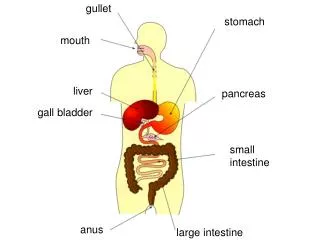
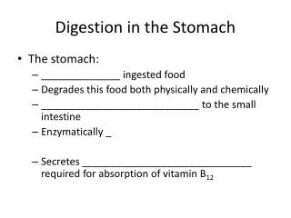
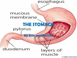
The Stomach. The normal gastric mucosa. Cardia – mainly mucus-secreting cells Fundus (body) – acid producing parietal cells, pepsin producing chief cells Pylorus – hormone (gastrin) production. Function of stomach. Mixing of food with acid/pepsin

The Stomach
E N D
Presentation Transcript
The normal gastric mucosa • Cardia – mainly mucus-secreting cells • Fundus (body) – acid producing parietal cells, pepsin producing chief cells • Pylorus – hormone (gastrin) production
Function of stomach • Mixing of food with acid/pepsin • Unique acid environment requires functional gastric surface mucus barrier, bicarbonate buffering and epithelial integrity
Congenital disorders • Hiatus hernia • Diaphragmatic hernia (through a non-physiological defect) • Congenital pyloric stenosis. Male infants with hypertrophy of pyloric smooth muscle leading to projectile vomiting
Gastritis • Acute gastritis often due to chemical injury (alcohol drugs) • Chronic gastritis: • Helicobacter pylori infection • Chemical damage (bile reflux, drugs) • Autoimmune (associated with vitamin B12 malabsorption (pernicious anaemia)
Acute gastritis • Drugs (non-steroidal anti-inflammatory drugs NSAID), alcohol cause acute erosion (loss of mucosa superficial to muscularis mucosae). Can result in severe haemorrhage • Acute Helicobacter infection has a prominent neutrophil infiltrate
Chronic gastritis ABC • A – autoimmune • B – bacterial (helicobacter) • C - chemical
Autoimmune chronic gastritis • Autoantibodies to gastric parietal cells • Hypochlorhydria/achlorhydria • Loss of gastric intrinsic factor leads to malabsorption of vitamin B12 with macrocytic,megaloblastic anaemia
Morphology of chronic gastritis • Chronic inflammatory cell infiltration • Mucosal atrophy • Intestinal (goblet cell) metaplasia Seen in Helicobacter and autoimmune gastritis (not chemical)
Helicobacter pylori • Adapted to live in association with surface epithelium beneath mucus barrier • Causes cell damage and inflammatory cell infiltration • In most countries the majority of adults are infected
Helicobacter gastritis • Acute inflammation mediated by complement and cytokines • Polymorphisms infiltrate epithelium and may be partly responsible for its destruction • An immune response is also initiated (antibodies may be detected in serum)
Helicobacter gastritis • 2 patterns of infection • Diffuse involvement of body and antrum (“pan gastritis” associated with diminishing acid output) • Infection confined to antrum (antral gastritis, associate with increased acid output)
Chemical gastritis • Commonly seen with bile reflux (toxic to cells) • Prominent hyperplastic response (inflammatory cells scanty) • With time – intestinal metaplasia
Consequences of gastritis • Peptic ulcer disease (Helicobacter) • Adenocarcinoma (all types) The “African enigma” – are complications of H.pylori infection less frequent in Africans? • Case not yet resolved
Peptic ulcer disease • A surface breach of mucosal lining of GI tract occurring as a result of acid and pepsin attack • Sites: • Duodenum (DU) • Stomach (GU) • Oesophagus • Gastro-enterostomy stoma • Related to ectopic gastric mucosa (e.g. in Meckel’s diverticulum)
Acute peptic ulcer • Like acute erosion but breaching muscularis mucosae • Specific examples • Curling’s ulcer (following severe burns) • Cushing’s ulcer (following head injury)
Chronic peptic ulcer • Complex epidemiology • DU most common in Europe, GU in Japan • Incidence of DU declining, GU stable
Pathogenesis • In normal acid/pepsin attack is balanced by mucosal defences • Increased attack by hyperacidity • Weakened mucosal defence – the major factor (H. pylori related)
Acid production • Tends to be high in DU patients. Antral gastritis causes increased gastrin production and acid secretion • Acid stimulates development of gastric metaplasia in the duodenum • Helicobacter organisms colonise the metaplastic epithelium and cause inflammatory damage leading to ulceration
Acid in GU • Pan gastritis diminishes acid secretion • Ongoing gastritis and epithelial damage is the main causal factor for ulceration
Helicobacter factors in pathogenesis • Some strains are more pathogenic than others. The Cag A (cytotoxic) antigen is one important virulence factor • Human variability also plays a part (e.g. individuals who produce high levels of IL-1b in inflammation get pan gastritis and GU, lower levels associated with antral gastritis and DU)
Morphology of peptic ulcers • Clean, non-elevated edge • Granulation tissue base (floor) • Underlying fibrosis
Complications of peptic ulcer • Perforation leading to peritonitis • Haemorrhage by erosion of vessel in base • Penetration of surrounding organ (liver/pancreas) • Obstruction (by scarring) – pyloric stenosis • (Cancer – rare event in true peptic ulcer)
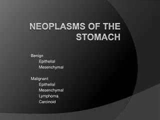
Gastric neoplasms • Polyps are common but usually not neoplastic (hyperplastic polyps. Hamartomas, ectopic pancreas) • Adenomas occur but are rare
Carcinoma of the stomach • The second most common fatal malignancy in the world • (after lung cancer) • Commonest in Far East (Japan) • Incidence declining • High mortality unless disease detected early
Pathology • Vast majority are adenocarcinomas • Arise on background of chronic gastritis, intestinal metaplasia, dysplasia • Most cases advanced at presentation
Macroscopic Pathology • Gross types • Polypoid • Ulcerative • Infiltrative (extreme is linitis plastica – “leather bottle stomach)
Microscopy • Intestinal type (forms glands – like cancers of colon and oesophagus) • Diffuse type – dissociated tumour cells often containing a mucinous “blob” – signet ring cells
Spread of gastric carcinoma • Local infiltration (through wall of stomach to peritoneum, pancreas etc) • Lymphatic – local and regional lymph nodes • Blood – liver, lungs • Transcoelomic (across peritoneal cavity). Often involves ovaries (esp. signet ring cancer) – Krukenberg tumour.
Less common gastric neoplasms • Lymphoma • Gastrointestinal stromal tumour (GIST) • Neuroendocrine (carcinoid) tumours
Gastric lymphoma • Malignant neoplasm of mucosa associated lymphoid tissue (MALT) • A (usually) low grade B-cell (marginal cell) lymphoma
Gastric lymphoma (maltoma) • Neoplastic cells infiltrate the epithelium (lymphoepithelial lesions) • Strongly associated with H. pylori and can be cured by eliminating infection.
Gastrointestinal stromal tumours (GIST) • Mesenchymal neoplasms • Derived from interstitial cells of Cajal (pacemaker cells controlling peristalsis) • Overexpress c-kit oncogene • Used as diagnostic aid on tissue • A target for therapy with tyrosine kinase inhibitor imatinib (also used in CML)
GIST • Larger tumours with high mitotic rate tend to behave malignantly • Stomach is commonest site
Neuroendocrine tumours • Carcinoids are tumours of resident neuroendocrine cells in gastric glands • Usually seen in context of chronic atrophic gastritis (driven by gastrin) • Clinical behaviour variable