Download
1 / 62
740 likes | 2.09k Vues
Use Of Electroencephalography (EEG) In The Management Of Seizure Disorders. Dr Lim Shih Hui Senior Consultant Neurologist Singapore General Hospital. Electroencephalography. 1st used in humans by Hans Berger in 1924 (the first report was published in 1929)

E N D
Use Of Electroencephalography (EEG) In The Management Of Seizure Disorders Dr Lim Shih Hui Senior Consultant Neurologist Singapore General Hospital
Electroencephalography • 1st used in humans by Hans Berger in 1924 (the first report was published in 1929) • A tracing of voltage fluctuations versus time recorded from electrodes placed over scalp in a specific array • Represent fluctuating dendritic potentials from superficial cortical layers • Required amplification • Deep parts of the brain are not well sampled
Types of EEG Recording • Routine • analog, digital • with computerized analysis & brain electrical activity mapping • Long-term Monitoring
Routine EEG Techniques • 20-min or longer sampling of brain activity • Written out or recorded directly on magnetic tape or digitally by computer • Disc electrodes are applied according to 10-20 system of electrode placement • Montages: referential, bipolar, changeable with digital recording
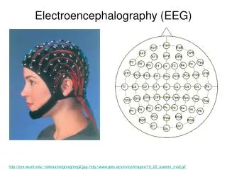
International 10-20 System of Electrode Placement • Established in 1958 • Electrodes are spaced at 10% or 20% of distances between specified anatomic landmarks • Use 21 electrodes, but others can be added • increase spatial resolution • record from specific areas • monitor other electrical activity (e.g. ECG, eye movements) • Odd number electrodes over left and even number over right hemisphere
Routine EEG Techniques • 20-min or longer sampling of brain activity • Written out or recorded directly on magnetic tape or digitally by computer • Disc electrodes are applied according to 10-20 system • Montages: bipolar, referential, changeable with digital recording
Routine Eye opening and closure Hyperventilation Intermittent photic stimulation 1, 5, 10, 15 & 20 Hz eyes open eyes closed eyes closure Optional Sleep deprivation Sedated sleep Specific methods of seizure precipitation video games visual patterns AED withdrawal Activations
Is a measure of brain function; supplement neuroimaging studies Provides direct rather than indirect evidence of epileptic abnormality May be the only test that shows abnormalities in epileptic patients Provides some spatial or localization information Low cost Low morbidity Readily repeatable Portable / ambulatory Strength and Advantages of EEG
Limitations and Disadvantages Of EEG • Detects cortical dysfunction but rarely discloses its etiology • Relatively low sensitivity and specificity • Subject to both electrical and physiologic artifacts • Influenced by state of alertness, hypoglycaemia, drugs • Small or deep lesions might not produce an EEG abnormality • Limited time sampling (for routine EEG) and spatial sampling • May falsely localize epileptogenic zone
Uses Of EEG In The Management of Seizure Disorders • To support a clinical diagnosis of epilepsy • To help to classify seizures • To help localize epileptogenic focus, especially in presurgical candidates • To quantify seizures • To aid in the decision of whether to stop AED treatment • Not a good guide to the effectiveness of treatment, except in absence seizures
Analyzing EEG Activities • Morphology • Distribution • Frequency • Voltage • Duration • State of the patient • Background from which activity is arising from • Similarity or dissimilarity to the other ongoing background rhythms
Guidelines To EEG Interpretation • Each EEG should be read with maximum possible objectivity • Ideally an EEG’er should describe the findings and make an EEG diagnosis without knowledge of the patient's history • Clinical significance of the findings can then be judged by integrating the EEG diagnosis with the history
EEG Interpretation • Normal • Lack of Abnormality • Abnormal • Non-epileptiform Patterns • Epileptiform Patterns
Epileptiform Patterns on Scalp-recorded EEG • Interictal Epileptiform Pattern • Electrographic Seizure Pattern • Isomorphic seizure pattern • Metamorphic seizure pattern
Criteria For Potentially Epileptogenic Transients • Clearly of cerebral and not artifactual origin • Abnormal for the age and the state of the patient • Have a significant epileptiform character and not one of the benign epileptiform variants
Physiologic Activities That Can Be Confused With Epileptiform Activities • Vertex transients of light sleep • Hypnagogic hypersynchrony • Positive occipital sharp transients of sleep (POST) • Mu rhythm • Lambda waves • Breach rhythms
Benign Variants Of Unknown Clinical Significance • Benign epileptiform transients of sleep (small sharp spikes) • 6- and 14-Hz positive spikes • Wicket spikes • Psychomotor variants (rhythmic mid-temporal theta discharge of drowsiness) • Subclinical rhythmic EEG discharge of adults • Phantom spike and wave
FP1 – F7 F7 – T3 T3 – T5 T5 – O1 FP2 – F8 F8 –T4 T4 – T6 T6 – O2 FP1 – F3 F3 – C3 C3 – P3 P3 – O1 FP2 – F4 F4 – C4 C4 – P4 P4 – O2 Fz – Cz Cz – Pz EKG PHOT Wicket Spikes
Examples Of Inter-ictal Epileptiform Patterns • Spikes • Sharp waves • Benign Epileptiform Discharges of Childhood • Spike-and-wave complexes • 3Hz Spike-and-wave complexes • Slow spike-and-wave complexes • Hypsarrhythmia • Photo-paroxysmal response
Interictal Spikes / Sharp Waves • Spikes (<70 msec in duration) or Sharp Waves (70-200 msec in duration) • Usually surface negative; occasionally bipolar or only surface positive • Monophasic, biphasic or polyphasic • Occur alone or accompanied by an after-coming slow wave (usually surface negative and higher in amplitude than the spike or sharp wave) • Occurs singly or in burst, lasting at most a few seconds • Focal or generalized • No clinical manifestation
Idiopathic Epilepsies Generalized 3 Hz spike-and-wave Polyspikes Atypical spike-and-wave Partial / Focal Benign focal epilepsy of childhood with centrotemporal spikes Benign focal epilepsy of childhood withoccipital spikes Symptomatic Epilepsies Generalized Hypsarrhythmia Slow spike-and-wave Paroxysmal fast activity Multiple independent spike foci Partial / Focal Temporal Frontal Centro-parietal Occipital Midline Inter-ictal Epileptiform Patterns
Fp1 – F7 F7 – T3 T3 – T5 T5 – O1 Fp2 – F8 F8 – T4 T4 – T6 T6 – O2 Fp1 – F3 F3 – C3 C3 – P3 P3 – O1 Fp2 – F4 F4 – C4 C4 – P4 P4 – O2 Fz – Cz Cz – Pz T1 – T2 A1 – A2 EKG Photic Benign Epileptiform Discharges of Childhood
Focal Inter-ictal Epileptiform Pattern • Temporal, frontal, occipital, centroparietal, centrotemporal, or midline • Relative frequency (Gibbs and Gibbs, 1952) • 1396 patients; Temporal: 73%; Frontal: 0.8% • Likelihood of seizures (Kellaway, 3526 children) • Temporal : 90-95% (91%) • Frontal : 70-80% (75%) • Parieto-occipital : 40-50% (48%) • Central : 30-40% (38%) • Focal spikes in a patient with a history of epileptic seizures indicates that the patient is likely to have focal or localization-related epilepsy syndrome
Epileptiform Activity In PeopleWithout Epilepsy • Zivin and Ajmone-Marsan, 1980 • 142/6497 (2.2%) of non-epileptic patients had IEDs • only 20/142 (14.1%) eventually developed epilepsy • Eeg-Olofsson et al, 1971 • 2.7% and 8.7% of 743 normal children had IEDs during wakefulness and sleep, respectively • Cavazutti et al, 1980 • 131/3726 children (3.5%) had IEDs on awake EEG • only 7/131 (5%) eventually developed seizures • Presence of interictal epileptiform discharges (IEDs) is not diagnostic of epilepsy
Normal EEG In People With Epilepsy • I know my patient has epilepsy. How can the EEG be normal? • If the EEG is normal, am I wrong in thinking that my patient has epilepsy?
Normal EEG In People With Epilepsy • Ajmone-Marsan & Zivin, 1970 • only 56% of 1824 EEGs from 308 patients (1-64 years) with known seizures showed IEDs on first EEG • IEDs recorded in another 26% in subsequent records • Holmes, 1986 • 25% of 24 paediatric patients with documented seizures on long-term monitoring had no IEDs • first and only EEG abnormalities recorded was complex partital seizures • Patients with well-documented seizures may have normal EEGs
Factors Responsible For Detection Of Epileptiform Discharges • Characteristic of Generator Source • Voltage of cortical discharge which is directly related to size/area of cortex involved in generation of synchronous activity • Distance between electrodes and the generator source • Orientation of dipole • Sampling Time • Activation
Factors Which Modify Spike Frequency • Sleep • Photic stimulation • Hyperventilation • Temporal relation to a seizure • Age of patient • Effect of anticonvulsant withdrawal
Recording of Focal Interictal Spikes • Yield of recording focal interictal spikes increases • during NREM sleep, especially stage 3/4 • after sleep deprivation • after seizures • during long-term monitoring • ? using supplementary electrodes (e.g. sphenoidal) • Occurrence of focal interictal spikes is not affected • by increasing or decreasing the AED dosages or level • by hyperventilation or photic stimulation • before seizures