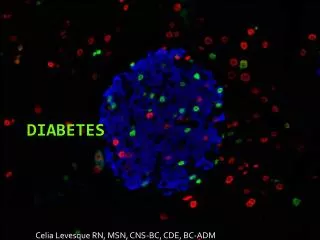
Langerhans’ Cell Histiocytosis
Langerhans’ Cell Histiocytosis. Chris Restrepo. Introduction and Background. Sometimes referred to as histiocytosis X LCH is a disease of abnormal clonal proliferation of a unique type of cell in the monocyte-macrophage cell line known as the Langerhans cell.

Langerhans’ Cell Histiocytosis
E N D
Presentation Transcript
Langerhans’ Cell Histiocytosis Chris Restrepo
Introduction and Background • Sometimes referred to as histiocytosis X • LCH is a disease of abnormal clonal proliferation of a unique type of cell in the monocyte-macrophage cell line known as the Langerhans cell. • It is named after the medical student Paul Langerhans, the first scientist to describe the cell (1868). Paul went on to be a German pathological anatomist
Introduction and Background • Peak incidence in infants 1-2 yo • Males X2 > females • Focal or systemic
Introduction and Background • Eosinophilic Granuloma (80%) • Localized benign form • Isolated to bone • Hand-Schuller-Christian disease (15-20%) • Skull lesions • Exopthalmos • Diabetes Insipidus • Letterer-Siwe disease (>10%) • Disseminated lesions involving multiple visceral organs
Pathogenesis • Exact cause is not completely understood. • Thought to possibly be due to… • disorder of the immune system • viral or other infectious cause • neoplasia
Pathology of Bone Lesions • Eosinophilic Granuloma • Most differentiated form • Best prognosis • Numerous eosinophils and Langerhans cells • May be asymptomatic
Pathology of Bone Lesions • Hand-Schuller-Christian Disease • Multiple bone lesions mainly located in the skull • Variable prognosis • May involve vertebral bodies leading to collapse
Pathology of Bone Lesions • Letterer-Siwe disease • Most agressive form • Children usually present with fever, skin lesions, hepatosplenomegaly, anemia, or lymphadenopathy • Usually in children under 2yo
Patient Presentation • Patients with osseous inolvement usually have pain, swelling, and a soft tissue mass • Prediliction for flat bones (esp the petrous ridge of the temporal bone) • 1/3 involve long bones (femur) • In long bones the diaphysis is involved 58% of the time
Plain Radiography • Acute Phase • Lesions can appear over a couple weeks • Aggressive looking lytic lesions with poorly defined margins • Difficult to differentiate from malignancy • Chronic Phase • Usually located in flat bones • Better defined • Early spontaneous healing may lead to reactive sclerosis
Plain Radiography • Long Bones • Begins with small area of medullary destruction • May regress to a well defined lesion with sclerotic margins • May progress to cause endosteal scalloping, cortical erosion, periosteal reaction, and soft tissue production • Chest wall lesions • Lytic and may be expansible
Plain Radiography • Skull Lesions • Oval-shaped and well defined • Look like punched out lytic lesions • Lytic lesions may contain a fragment of intact bone referred to as a button sequestrum • Skull may take on “geographic” appearance • NO periosteal reaction or reactive sclerosis • Spinal Lesions • Predilection for thoracic spine • May lead to near collapse with “vertebra plana” appearance
Scintigraphy • After Dx, further investigation involves detection of other bony lesions by a radiographic skeletal survey or bone scintigraphy • Some suggest to do both • Conway • 19% were missed on bone scan • 29% were missed on plain radiographs • Radiography is more cost-effective but scintigraphy is probably more sensitive
Computed Tomography • Helps confirm presence of bone lesion as well as demonstrating any cortical destruction and soft tissue involvement • Preservation of disc space above and below vertebral body helps differentiate lesion from osteomyelitis • May use CT-guided Bx to establish dx of LCH in the spine • May detect very small lesions and rare cortical or sacral lesions
Magneti Resonance Imaging • Very sensitive but findings are nonspecific • Can easily detect extension of lesions into the dura matter or brain • Lytic lesions are hypointense on T1 and hyperintense on T2 • Healing shows a decrese in signal intensity on T2
Differential Diagnosis • Destructive lesion – malignancy such as Ewing Sarcoma, lymphoma, leukemia, and mets as well as osteomyelitis • MR imaging findings are nonspecific and can be confused with Ewing Sarcoma, Osteoblastoma, and Osteomyelitis • Vertebral lesions can be confused with lymphoma-leukemia, mets, and trauma.