Download

1 / 25
260 likes | 937 Vues
0. Orientation Lab Safety and Restriction Enzymes. Kabi Neupane, Ph.D. Leeward Community College. 0. Objectives. Familiarize with laboratory safety Learn about Restriction enzymes Perform a restriction digestion of Arabidopsis genomic DNA. 0. Laboratory Safety. Emergency procedures

E N D
0 Orientation Lab Safety and Restriction Enzymes Kabi Neupane, Ph.D. Leeward Community College
0 Objectives • Familiarize with laboratory safety • Learn about Restriction enzymes • Perform a restriction digestion of Arabidopsis genomic DNA
0 Laboratory Safety • Emergency procedures • Eye wash stations • Locate both eye wash stations • Personal safety • Lab coats, gloves, goggles • Chemical safety • Material Safety Datasheets (MSDS) • Red binder located on the back • Biological safety
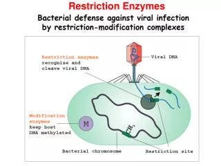
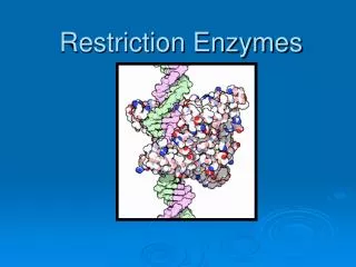
0 Enzymes • Enzymes are proteins • biological catalysts help drive biochemical reactions • Enzyme names end with an ase(eg., endonuclease) • Bacteria have evolved a class of enzymes that destroy foreign DNA (eg. Virus DNA). • protect bacteria from bacteriophages (Viruses). • Bacteriophages cannot multiply if their DNA is destroyed by the host.
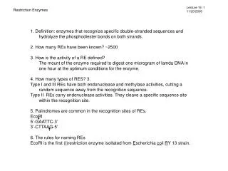
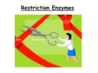
0 Restriction Endonucleases • Restriction endonucleases RESTRICT viruses • Viral genome is destroyed upon entry • Restriction endonuclease = Restriction enzymes • Endo (inside), nuclease (cuts nucleic acid) • Restriction endonuclease recognizes a short and specific DNA sequence and cuts it from inside. • The specific DNA sequence is called recognition sequence
0 Discovery • 1952-53: Luria and Human discovered the phenomenon of restriction and modification • Named as host-induced, or host-controlled, variation.
0 Bacteriophage Life Cycle http://student.ccbcmd.edu/courses/bio141/lecguide/unit3/viruses/lytsum.html
0 Restriction? • Bacteriophages varied in their ability to grow on different strains of E.coli. • Once growth was achieved on one host strain, the phages could continue to grow happily on this strain. • However, the phages were now restricted in their ability to grow on other strains.
0 Nomenclature • Smith and Nathans (1973) proposed enzyme naming scheme • three-letter acronym for each enzyme derived from the source organism • First letter from genus • Next two letters represent species • Additional letter or number represent the strain or serotypes • For example. the enzyme HindII was isolated from Haemophilus influenzae serotype d.
0 Few Restriction Enzymes
0 Classification • Synonymous to Restriction Endonuclease • Endonuclease: Cut DNA from inside • Highly heterogeneous • Evolved independently rather than diverging form a common ancestor • Broadly classified into four Types
0 R-M System • Restriction-modification (R-M) system • Endonuclease activity: cuts foreign DNA at the recognition site • Methyltransferase activity: protects host DNA from cleavage by the restriction enzyme. • Methyleate one of the bases in each strand • Restriction enzyme and its cognate modification system constitute the R-M system
0 Protection of Self DNA • Bacteria protect their self DNA from restriction digestion by methylation of its recognition site. • Methylation is adding a methyl group (CH3) to DNA. • Restriction enzymes are classified based on recognition sequence and methylation pattern.
0 Type I • Multi-subunit proteins • Function as a single protein complex • Contain • two R (restriction) subunits, • two M (methylation) subunits and • one S (specificity) subunit • Cleave DNA at random length from recognition site
0 Type III • Large enzymes • Combination restriction-and-modification • Cleave outside of their recognition sequences • Require two recognition sequences in opposite orientations within the same DNA molecule • No commercial use or availability
0 Type IV • Cleave only modified DNA (methylated, hydroxymethylated and glucosyl-hydroxymethylated bases). • Recognition sequences have not been well defined • Cleavage takes place ~30 bp away from one of the sites. • Sequence similarity suggests many such systems in other bacteria and archaea.
0 Type II • Most useful for gene analysis and cloning • More than 3500 REs • Recognize 4-8 bp sequences • Need Mg 2+ as cofactor • Cut in close proximity of the recognition site • Homodimers • ATP hydrolysis is not required
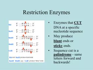
0 Recognition Sequences • Each restriction enzyme always cuts at the same recognition sequence. • Produce the same gel banding pattern (fingerprint) • Many restriction sequences are palindromic. For example, (Read the same in the opposite direction (eg. madam, race car…) • 5’ GAATTC 3’ • 3’ CTTAAG 5’
5’ P - - OH 3’ HindIII - P 5’ 3’ OH - EcoRI 0 Sticky End Cutters • Most restriction enzymes make staggered cuts • Staggered cuts produce single stranded “sticky-ends” • DNA from different sources can be spliced easily because of sticky-end overhangs.
AluI HaeIII 0 Blunt End Cutters • Some restriction enzymes cut DNA at opposite base • They leave blunt ended DNA fragments • These are called blunt end cutters
0 Restriction Enzyme Use • Discovery of enzymes that cut and paste DNA make genetic engineering possible. • Restriction enzyme cuts DNA and generates fragments • Ligase joins different DNA fragments • DNA fragments from different species can be ligated (joined) to create Recombinant DNA
0 Cloning Vectors Play
0 Typical Restriction Digest Sterile, deionized water 16.3 µl RE 10X Buffer 2.0 µl Acetylated BSA, 10µg/µl 0.2 µl DNA, 1µg/µl 1.0 µl Mix by pipetting, then add: Restriction Enzyme, 10u/µl 0.5 µl Final volume 20.0 µl
0 How does it Look after Restriction Digestion? Genomic DNA Digest Plasmid DNA Digest
0 Questions?