Download
1 / 28
320 likes | 615 Vues
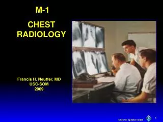
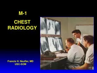
Intro to Chest Radiology. Develop a System. Helps you remember things to check Mneumonic vs anatomic. Basics. Technique PA & Lateral AP or Portable Indication Identification Rotation Penetration Inspiration Comparison. PA & Lateral More information Two views Standardized Distance

E N D
Develop a System • Helps you remember things to check • Mneumonic vs anatomic
Basics • Technique • PA & Lateral • AP or Portable • Indication • Identification • Rotation • Penetration • Inspiration • Comparison
PA & Lateral More information Two views Standardized Distance Pt needs to be stable Portable Quick Anywhere One shot No standardization Techniques
Basics • Indication • The more information, better read • Identification of patient • Penetration • See vertebral bodies through the heart? • Rotation • Clavicular heads in relation to vertebral spine • Inspiration • Count 8-9 posterior ribs
Things to see • ABCDE… • Airways • Trachea, endotracheal tube, etc • Bones • Clavicles, ribs, etc… • Cardiac • Diaphragm (Right hemidiaphragm slightly higher (~1.5 cm) • Everything else (tubes), effusions
Darker areas radiolucent Pneumothorax Cysts/bulla Air bronchograms Lighter areas Opacities “infiltrates” Blood Pus Water Nodules or mass Lung findings
Opacities • Lobar or not…. • Pneumonia • Pulmonary Edema • “fluffy,” diffuse, “bat wing” distribution • Hemorrhage • Cant tell by xray, need bronch
Next Step… • COMPARE, COMPARE, COMPARE… • New or old • Any growth • Pattern of calcification • Next Imaging step vs. other test
Summary • ABCDE… • Point out abnormalities • Compare to old films • Give your impression