Download
1 / 47
500 likes | 2.26k Vues
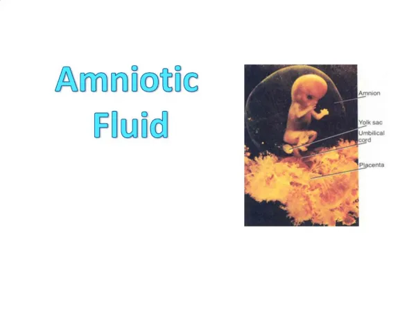
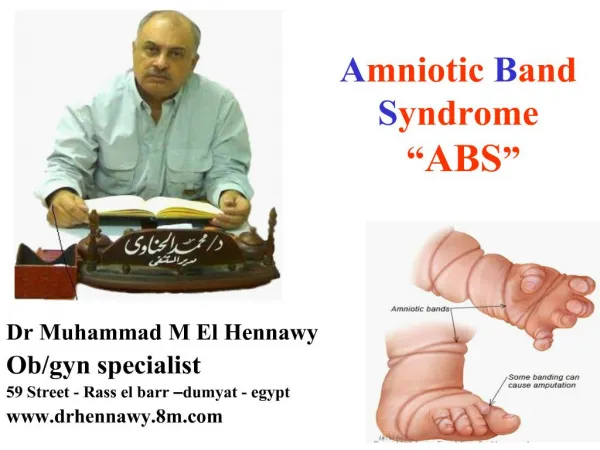
What is Amniotic Band Syndrome?. It is a set of congenital birth defects believed to be caused by entrapment of fetal parts (usually a limb or digits) in fibrous amniotic bands while in utero. . Typs Of ABS. A membrane formed at an early stage of pregnancy may cause severe damage to the structure of the various tissues, resulting in deformities in a large number of organs

E N D
1. Amniotic Band Syndrome �ABS� Dr Muhammad M El Hennawy
Ob/gyn specialist
59 Street - Rass el barr �dumyat - egypt
www.drhennawy.8m.com
3. Typs Of ABS A membrane formed at an early stage of pregnancy may cause severe damage to the structure of the various tissues, resulting in deformities in a large number of organs � this often results in intrauterine death. Defects in these cases usually include defects in the location and structure of the heart, omphalocele, gastroschisis, anencephalus, cleft face, hydrocephalus, etc.
4. Amniotic Band Syndrome - Alternative Names Some of these are different names for ABS and some are other syndromes that are often confused with ABS.� Misdiagnosis is apparently common.�
�
Amniotic Deformity, Adhesions and Mutilations
Amniotic band disruption complex or sequence
Amniotic bands and sheets
Annular constriction bands�
Congenital ring constriction�
Constriction Band Syndrome� and Amniotic Constriction Band Syndrome
Intrauterine amputation
Streeter's dysplasia
TEARS (The Early Amnion Rupture Spectrum)
Congenital Transverse Defects or Deficiency
Limb-body Wall Complex�
Amniotic deformity adhesions mutilations (ADAM).
ADAM Complex
Amniotic band sequence,
Amniochorionic mesoblastic fibrous strings
Congenital amputation,
Congenital constricting bands
Tissue bands
�
5. Incidence Amniotic band syndrome (ABS) is an uncommon fetal malformation with increasing prevalence
Amniotic banding affects approximately 1 in 1,200 to 1 in 15,000 live births.
It is also believed to be the cause of about 178 in 10,000 miscarriages.
About 80% of cases involve the hands and fingers and a significant number of clubfoot cases are correlated with ABS
6. Prenatal risk factors No distinct sex predilection has been determined.
Nearly sixty percent of the cases have some sort of abnormal gestation history.
Prenatal risk factors associated with amniotic band syndrome include prematurity (<37 weeks), low birth weight (<2500 g), maternal illness (during pregnancy), maternal drug exposure and maternal hemorrhage/trauma.
7. Causes Amniotic Band Syndrome is not genetic (i.e. not inherited).
It is extremely unlikely that ABS will affect a future pregnancy.�
To date, no prenatal factors have been associated with ABS
The primary event is a rupture of the amniotic membrane and its detachment from the chorion with amniotic fluid leaking through the tear (spontaneously or after trauma to the lower abdomen ).
As a result, the fetus can move digits or limbs through this tear and exit the amniotic cavity (partially or completely).
The outer surface of the amnion, and to a lesser degree the naked chorion, produce mesodermic fibrous strings which may entangle and entrap different fetal organs, leading to constriction and amputation anomalies.
These deductions agree with the increased frequency of constriction bands located more distally on the digits, hands and feet, as well as with the varying severity of constrictions, ranging from slight grooves in the skin to actual amputation of the digit or limb
However, this theory does not explain the association of amniotic bands with damage to internal organs and other severe anomalies.
8. The Timing Of The Rupture The timing of the rupture is believed to occur between 28 days after conception to 18 weeks of gestation.
Late bands can occur and present at birth, even after a normal ultrasound was performed earlier in the pregnancy.
9. Features or Stages The constriction of appendages by amniotic bands may result in:
Restrictions of the growth around the digits, arms and legs
Constriction rings around the digits, arms and legs - the bands will partially constrict the blood flow in the affected appendage causing a deep groove in the baby's skin
Swelling of the extremities distal to the point of constriction (congenital lymphedema)
Amputation of digits, arms and legs (congenital amputation)
10. Range of effects
The defect could be merely cosmetic, depending on the severity of the constriction.
Deeper bands may cause lymphatic obstruction leading to edema and vascular compromise, conditions that require immediate release.
Abnormalities may occur distal to the constriction, such as anterolateral bowing, hemihypertrophy, pseudarthrosis, leg-length discrepancy, and teratologic clubfeet. These conditions may lead to limited function and movement.
Early in gestation, spontaneous abortions may result from the encircling bands. If the constriction occurs after development is almost complete, fissures, acrosyndactylization, or intrauterine amputation occur typically on the extremities.
11. ABS affects the hands in almost 90% of cases. The distal portion of the extremities is most often involved, especially in the longer central fingers of the hand. The thumb and small finger are rarely involved, presumably because of their shorter lengths. In the feet, constricting bands most commonly involve the big toe (hallux).
Lymphatic and vascular compromise may result from severe band compression. Immediate surgical release is required if at birth the child presents with a swollen, engorged digit or limb.
More often, the constricted digit or limb has been amputated in utero. Acrosyndactyly occurs after digital separation is complete, but the fingers get twisted by bands and eventually join together.
12. Clubfoot occurs in up to 25% of cases of amniotic banding. In half of occurrences of clubfoot, a tight band wraps around the peroneal nerve, which causes muscle imbalance and clubfoot.
Constriction bands across the face and head may result in facial clefts. Cleft lip and palate require reconstruction when the child is approximately 3 to 6 months of age.
13. ABS may affect the face with cleft lip or palate, asymmetric microphthalmia or severe nasal deformity. Encephalocele may be a manifestation of Amniotic Band Syndrome / ABS, especially when eccentrically placed off the midline.
Abdominal wall defects - typically large defects with free-floating intestine but large enough for the lines to herniate outside the abdomen, can also be the result of Amniotic Band Syndrome / ABS.
The characteristic appearance of an aberrant sheet or band of amnion attached to the fetus with resultant deformity and restriction of motion allows a diagnosis of Amniotic Band Syndrome / ABS to be made. Prenatal diagnosis is the exception rather than the rule.
14. The findings in Amniotic Band Syndrome / ABS may be limited to isolated defects, including isolated facial cleft, digital amputation or mild elephantiasis of an extremity beyond a constrictive band. These features may be difficult to diagnose using ultrasound because the detailed fetal visualization required is beyond the scope of routine obstetrical ultrasound examinations.
At the worst end of the spectrum, the fetus may be so severely deformed by the amniotic bands that the spine is contracted and organs are formed in perplexing and bizarre proportions. The head may be completely misshapen or absent.
The bands responsible for these deformities are rarely seen and a presumptive diagnosis of Amniotic Band Syndrome / ABS is made based on the commonly associated deformities.
15. The spinal deformities in Amniotic Band Syndrome can be severe, manifesting as kyphotic lordosis or scoliosis as well as severe rotational abnormalities, even spinal amputation. While spinal deformity can be seen in other syndromes, severe spinal deformity should suggest Amniotic Band Syndrome / ABS.
Spinal deformity associated with an abdominal-wall defect is particularly suggestive of Amniotic Band Syndrome.
While the typical appearance of an omphalocele is possible, the more common defect is a large slash-like defect of both the thoracic and abdominal cavities with evisceration.
16. These defects are associated with exteriorized bowel, liver and sometimes heart without an enveloping membrane. When associated with limb abnormalities, this is characteristic of the limb-body-wall complex form of Amniotic Band Syndrome.
Deformation of the calvarium is another group of anomalies characteristic of Amniotic Band Syndrome.
If complete, the fetus may appear anencephalic. If partial, the fetus may appear to have an encephalocele.
17. How is ABS diagnosed? ABS is very difficult to diagnose.
Prenatal ultrasound may not be able to identify the bands as the individual strands are small and hard to see on ultrasound but only see the results of the fusion such as missing or deformed limbs.
Misdiagnosis is also common, up to 70% of all amniotic bands seen on prenatal ultrasound disappear upon repeat ultrasound. If the ultrasound demonstrates amniotic bands with free fetal movement and lack of fetal attachment to the band, they are termed innocent amniotic bands and pose little risk to the fetus.
3D ultrasound and MRI can be used for more detailed and accurate diagnosis of bands and the resulting damage/danger to the fetus.
18. DD Of Amniotic Sheetsfrom Amniotic Bands Amniotic sheets are the most common with an incidence of 0.6%. They are thought to be caused by scarring inside the womb or 'senechiae' from previous operations, such as D&C. As the membranes develop, they surround these sheets of scar tissue. Often they aren't seen until later on in pregnancy, presumably following rupture or compression by the growing baby. Amniotic sheets have been found to be associated with an increased risk of early labour.
Amniotic bands are even less common, affecting 1 in 1200 (0.08%) of all pregnancies. They are fibrous strands of membrane stretching from the outer membrane surface into the amniotic cavity. They are thought to originate when the inner membranes (amnion) rupture without injury to the outer membrane (chorion). The ruptured amnion remains as a plaque or fragments into bands which stretch across the chorionic cavity. Amniotic bands are thought to happen spontaneously or in association with trauma to the abdomen. There has been the suggestion of a relationship between amniotic bands, limb shortening and early chorion villous sampling (CVS).
19. Other Bands
Other less common types of band-like appearing structures may also be observed on obstetric sonogram.
These include: chorio-amniotic separation (normal finding in the 1st trimester up to 16 weeks), velamentous cord insertion, uterine fusion abnormalities (bicornuate, septate uterus, etc.), and remaining membranes after demise of a twin.
In these situations, correlation of ultrasound features with patient's clinical history can be useful.
20. Approach to Evaluating Band-like Structures The following diagram can be utilized to aid diagnosis when a band-like structure is visualized in the uterus. The authors advise caution, however, as there may be some overlap of appearances in the different categories
21. Ultrasound Appearance: Usually thin membrane-like strands criss-crossing the amniotic sac and attached to fetal body parts. (ABS)
22. First trimester transvaginal sonogram showing an interesting synechia
Ultrasound Appearances:Shelf-like thick band communicating along it's length with the uterine wall along it's length. In other words, a synechia has a base and a free edge. This appearance is caused by a combination of the fibrous synechia itself, and the complete wrapping of fetal membranes around the synechia. �
23. Prevention Amniotic band syndrome is considered an accidental event and it does not appear to be genetic or hereditary, so the likelihood of it occurring in another pregnancy is remote.
The cause of amnion tearing is unknown and as such there are no known preventative measures.
24. Interventions and Treatment Treatment usually occurs after birth and where plastic and reconstructive surgery is considered to treat the resulting deformity.
Plastic surgery ranges from simple to complex depending on the extent of the deformity. Physical and occupational therapy may be needed long term.
In rare cases, if diagnosed in utero, fetal surgery may be considered to save a limb which is in danger of amputation or other deformity. This typically would not be attempted if neither vital organs nor the umbilical cord are affected.
25. Fetal Treatment In utero limb salvage: fetoscopic release of amniotic bands for threatened limb amputation.
Fetoscopic laser release of amniotic bands in extremity ABS offers the potential to prevent limb amputation
A small camera is inserted through the mother�s abdomen and uterus into the amniotic sac in order to see and cut the bands.
Cutting the amniotic bands is performed utilizing a fetoscopic technique.
The procedure is typically performed under regional anesthesia.
Early fetoscopic release may prevent amputation and allow improved limb development.
26. Child Treatment Indications for intervention depend on the medical stability of the child and on the neurovascular status of the limb.
Bands that only cosmetically affect the superficial skin generally do not require any intervention.
Only the tight constriction bands, resulting in gross lymphedema, vascular compromise, or both necessitate immediate surgical release.
Surgery also is indicated for patients with syndactyly or acrosyndactyly that compromises hand function. Thumb amputation (which is rare), club feet, cleft lip, and cleft palate require reconstruction, but these procedures can be performed electively at a later time.
27. Special Considerations For Delivery Type of delivery - Typically, pregnancies with ABS do not require cesarean delivery. The need for fetal intervention should not impact the mode of delivery. The delivery plan will be carefully discussed.
Place of delivery - If all the prenatal monitoring suggests that the baby is doing well, the baby can be delivered at the hospital. However, the hospital should be prepared to handle any immediate needs of the newborn and have a neonatal intensive care unit with the capability to provide specialized care.
Time of delivery - Unless there are signs of serious complications of ABS, there is no reason to intentionally induce an early delivery. The team at the Center may recommend early delivery for pregnancies that appear to be in danger
28. Follow-up and prognosis
All patients with ABS should be monitored regularly until skeletal maturity is reached, because of the potential for recurrence of the rings and for secondary contractures that may develop
The prognosis is good for limbs affected by isolated superficial extremity bands. Aside from cosmetic irregularities, no functional defects are usually present. Deeper bands may be associated with progressive problems leading to lymphatic and neurovascular compromise that requires operative intervention.
For patients with acrosyndactyly, hand function is limited secondary to stiffness of the joints, but reconstruction can result in good prehension and grasp. Children whose limbs have been amputated in utero usually adapt well to their physical limitations, and aside from fitting with a prosthesis, little often needs to be done. In children with a transverse deficiency proximal to the ankle joint, a prosthesis is required for full function.
29. Prognosis The prognosis depends on the location and severity of the constricting bands.
Every case is different and multiple bands may be entangled around the fetus.
Bands which wrap around fingers and toes can result in syndactyly or amputations of the digits.
In other instances, bands can wrap around limbs causing restriction of movement resulting in clubbed feet.
In more severe cases, the bands can constrict the limb causing decreased blood supply and amputation.
Amniotic bands can also sometimes attach to the face or neck causing deformities such as cleft lip and palate.
If the bands become wrapped around the head or umbilical cord it can be life threatening for the fetus.
30. Acrosyndactyly Acrosyndactyly is a more complex type of syndactyly. The fingers had separated but a band formed around the fingers causing them to refuse during development.
These images are the left and right hand of a newborn.
31. Amputated Big Toe ABS is the most common cause of a congenital amputation of a limb/digit.
32. Banded Foot When bands wrap around limbs during development they can constrict the limb causing decreased blood supply and amputation.
33. Banding on Leg This image shows a banding line on newborn's leg.
34. Cleft Lip & Palate Oral clefting occurs when the tissues of the lip and/or palate of a fetus don't grow together early in pregnancy.
Children with clefts often don't have enough tissue in their mouths, and the tissue they do have isn't fused together properly to form the roof of their mouths.
35. The umbilical cord of the dead twin is completely amputated.
36. Clubfeet Photo A strong relationship between ABS and clubfoot exists.
A 31.5% of associated clubfoot deformity and ABS can be correlated with 20% occurring bilaterally.
37. Nubbins This is typical of Congenital Transverse Deficiency, a form of ABS.
A short below-the-elbow amputation. The fingers may be represented only by nubbins or dimpling found on the end.
38. Overlapping, Underlapping Toes Congenital Overlapping toes are characterized by one toe lying on top of an adjacent toe. Congenital Under lapping toes usually involve the fourth and fifth toes.
39. Syndactyly - at birth A hand at birth, the banding line from the wrist to the pinky finger and around the wrist. The three fingers are webbed together (syndactyly).
The three fingers are small, with abnormal finger nails, and missing some joints.
There is also some lymphedema (swelling at the joint) of the index finger middle joint.
40. ABS affecting the leg above ankle.
41. Left hand with constriction bands, dystrophic nails and fusion of digits II-V by amniotic membrane.
42. Right foot with stunted growth of all digits, dystrophic nails, constriction bands and partial syndactyly of digits II to V.
44. Clubfoot Treatment The accepted method of treating clubfeet is by the Ponseti method of Serial Plaster Casting. Treatment should be started right away. The initial treatment consists of manipulating the foot to get it to the best position possible, and then holding the correction in a cast.
45. Syndactyly - Surgically Separated This photo is after two operations that separated his webbed fingers (syndactyly).
46. Distraction Augmentation Manoplasty This surgery creates growth at the rate of an inch of bone a month, to create new palms and to lengthen the fingers. It consists of the insertion of a device through the bone which is a complex group of bars, nails and steel screws. It requires screwing several turns per day to reach a growth rate of up to 1 ml. daily.
47. Summary Amniotic Band Syndrome can be difficult to diagnose. Ultrasound does not usually reveal it.
Even though the incidence of the condition is very low, obstetricians need to be aware of the possibility of umbilical cord constriction by an amniotic band.
The presentation of a patient with decreased fetal heart rate and a history of amniocentesis must to be monitored closely. There is a high fetal death rate associated with this diagnosis. When the fetus is at a viable gestational age, immediate delivery may be indicated.
When Intrauterine Demise does occur, amniotic band syndrome should be included as one of the possible etiologies. Thorough evaluation of the placenta and cord is necessary to rule out constriction of the cord by Amniotic bands.
Although the finding of Amniotic Band Syndrome may not change the outcome identifying a definitive cause for the parents may provide a small measure of closure in the context of this tragic event.