Download
1 / 33
330 likes | 1.02k Vues
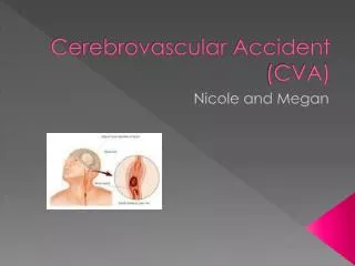
Cerebrovascular Accident Dr. Zolfaghari Assistant Professor of Emergency Medicine. Cerebrovascular Accident. Results from ischemia to a part of the brain or hemorrhage into the brain that results in death of brain cells . #1 leading cause of disability Third most common cause of death

E N D
Cerebrovascular AccidentDr. ZolfaghariAssistant Professor of Emergency Medicine
Cerebrovascular Accident • Results from ischemia to a part of the brain or hemorrhage into the brain that results in death of brain cells. • #1 leading cause of disability • Third most common cause of death • 25% with initial stroke die within 1 year • 25% will live with permanent disability • 50-75% will be functionally independent • Physical, cognitive, emotional, & financial impact
Cerebrovascular AccidentRisk Factors • Non modifiable: • Age – Occurrence doubles each decade >55 years • Gender – Equal for men & women; women die more frequently than men • Race – African Americans, Hispanics, Native Americans, Asian Americans -- higher incidence • Heredity – family history, prior transient ischemic attack, or prior stroke increases risk
Cerebrovascular AccidentRisk Factors Controllable Risks with Medical Treatment & Lifestyle Changes: High blood pressure DiabetesCigarette smoking TIA (Aspirin)High blood cholesterol ObesityHeart Disease Atrial fibrillation Oral contraceptive use Physical inactivity Sickle cell disease Asymptomatic carotid stenosisHypercoagulability
Cerebrovascular AccidentAnatomy of Cerebral Circulation • Blood Supply • 20% of cardiac output—750-1000ml/min • >30 second interruption– neurologic metabolism is altered; metabolism stops in 2 minutes; brain cell death < 5 mins.
Cerebrovascular AccidentAnatomy of Cerebral Circulation • Blood Supply • Anterior: Carotid Arteries – middle & anterior cerebral arteries • frontal, parietal, temporal lobes; basal ganglion; part of the diencephalon (thalamus & hypothalamus) • Posterior: Vertebral Arteries – basilar artery • Mid and lower temporary & occipital lobes, cerebellum, brainstem, & part of the diencephalon • Circle of Willis – connects the anterior & posterior cerebral circulation
Cerebrovascular AccidentPathophysiology • Atherosclerosis: major cause of CVA • Thrombus formation & emboli development
Cerebrovascular AccidentClassifications • Ischemic Stroke • Thrombotic • Embolic • Hemorrhagic Stroke • Intracerebral Hemorrhage • Subarachnoid Hemorrhage
Cerebrovascular AccidentPathophysiology • Border Zone: reversible area that surrounds the core ischemic area in which there is reduced blood flow but which can be restored (3 hours +/-)
CVA? - Call 115 • Sudden numbness or weakness of face, arm, or leg, especially on one side of the body. • Sudden confusion or trouble speaking or understanding speech. • Sudden trouble seeing in one or both eyes. • Sudden trouble walking, dizziness, or loss of balance or coordination • Sudden severe headache with no known cause.
Cerebrovascular AccidentClinical ManifestationsAnterior Cerebral Artery Involvement • Contralateral • weakness of proximal upper extremity • sensory & motor deficits of lower extremities • Urinary incontinence • Sensory loss (discrimination, proprioception) • Contralateral grasp & sucking reflexes may be present • Apraxia – loss of ability to carry out familiar purposeful movements in the absence of sensory or motor impairment • Personality change: flat affect, loss of spontaneity, loss of interest in surroundings • Cognitive impairment
Cerebrovascular AccidentClinical ManifestationsMiddle Cerebral Artery Involvement • Contralateral weakness • Hemiparesis; hemiplegia • Contralateralhemianesthesia • Loss of proprioception, fine touch and localization • Dominant hemisphere: aphasia • Nondominant hemisphere – neglect of opposite side; anosognosia – unaware or denial of neuro deficit • Homonymous hemianopsia – defective vision or blindness right or left halves of visual fields of both eyes
Cerebrovascular AccidentClinical ManifestationsPosterior Cerebral Artery & Vertebrobasilar Involvement • Alert to comatose • Unilateral or bilateral sensory loss • Contralateral or bilateral weakness • Dysarthria – impaired speech articulation • Dysphagia – difficulty in swallowing • Hoarseness • Ataxia, Vertigo • Unilateral hearing loss • Visual disturbances (blindness, homonymous hemianopsia, nystagmus, diplopia)
Cerebrovascular AccidentHemorrhagic Stroke • Hemorrhagic Stroke • 15% of all strokes • Result from bleeding into the brain tissue itself • Intracerebral • Subarachnoid
Cerebrovascular AccidentHemorrhage Stroke Intracerebral Hemorrhage • Rupture of a vessel • Hypertension – most important cause • Others: vascular malformations, coagulation disorders, anticoagulation, trauma, brain tumor, ruptured aneurysms • Sudden onset of symptoms with progression • Neurological deficits, headache, nausea, vomiting, decreased LOC, and hypertension • Prognosis: poor – 50% die within weeks • 20% functionally independent at 6 months
Cerebrovascular AccidentHemorrhagic-Subarachnoid • Hemorrhagic Stroke–Subarachnoid Hemorrhage • Intracranial bleeding into the cerebrospinal fluid-filled space between the arachnoid and pia mater membranes on the surface of the brain
Cerebrovascular AccidentHemorrhagic-Subarachnoid • Commonly caused by rupture of cerebral aneurysm (congenital or acquired) • Saccular or berry – few to 20-30 mm in size • Majority occur in the Circle of Willis • Other causes: Arteriovenous malformation (AVM), trauma, illicit drug abuse • Increases with age and more common in women
Cerebrovascular AccidentHemorrhagic-SubarachnoidCerebral Aneurysm • Warning Symptoms: sudden onset of a severe headache – “worst headache of one’s life” • Change of LOC, Neurological deficits, nausea, vomiting, seizures, stiff neck • Despite improvements in surgical techniques, many patients die or left with significant cognitivedifficulties
Hemorrhagic-SubarachnoidCerebral Aneurysm • Treatment: • Vasospasm prevention – Calcium Channel Blockers • Surgical Treatment
Cerebrovascular AccidentTransient Ischemic Attack • Temporary focal loss of neurologic function • Caused by ischemia of one of the vascular territories of the brain • Microemboli with temporary blockage of blood flow • Lasts less than 24 hrs – often less than 15 mins • Most resolve within 3 hours • Warning sign of progressive cerebrovascular disease
Cerebrovascular AccidentTransient Ischemic Attack • Diagnosis: • CT without contrast • Confirm that TIA is not related to brain lesions • Cardiac Evaluation • Rule out cardiac mural thrombi • Treatment: • Medications that prevent platelet aggregation • ASA, Plavix • Oral anticoagulants
Cerebrovascular AccidentGoals of prehospital Management • Immediate – assess & stabilize • ABCs, VS • Rapid Identification • Active stroke team • Rapid transport • Oxygen if hypoxic ( O2 sat < 95% ) • IV access • Check glucose • 12-lead EKG • Immediate Neuro Assessment • Establish symptom onset • Review hx • Stroke Scale, Facial droop; arm drift; abnormal speech
Cerebrovascular AccidentGoals of hospital Management • Immediate – reassess & stabilize • ABCs, VS • Rapid neurologic reassessment • Check glucose • Body temperature • Complication assessment • 12-lead EKG • Emergent CT scan of brain • BP management: • Fibrinolytic therapy • Specific medical condition
Cerebrovascular AccidentGoals for Management • CT Scan – No hemorrhage: • Consider Fibrinolytic therapy • Check for exclusions • tPA • No anticoagulants or antiplatelet therapy for 24 hours • If not a candidate: Antiplatelet Therapy • CT Scan – Hemorrhage: • Neurosurgery? • If no surgery: Stroke Unit • Monitor BP and treat Hypertension • Monitor Neuro status • Monitor blood glucose and treat as needed • Supportive therapy
Cerebrovascular AccidentTreatment Goals • Drug Therapy – Ischemic CVA – to reestablish blood flow through a blocked artery • Thrombolytic Drugs: tPA (tissue plasminogen activator) • Administered within 3 hours of symptoms of ischemic CVA • Confirmed DX with CT • Anticoagulant • Anti platelet