Download
1 / 40
540 likes | 1.27k Vues
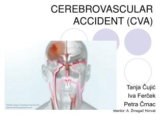
Cerebrovascular Accident (Stroke). Dayna Ryan, PT, DPT Winter 2012. CVA/Stroke Epidemiology. Population Statistics for 2006 Age 18-44……………………..0.5% 45-64……………………..2.4% 65-74……………………..7.6% 75 +………………………11.2% Sex Male ......................2.9% Female...................2.3%. Race

E N D
Cerebrovascular Accident (Stroke) Dayna Ryan, PT, DPT Winter 2012
CVA/Stroke Epidemiology Population Statistics for 2006 • Age • 18-44……………………..0.5% • 45-64……………………..2.4% • 65-74……………………..7.6% • 75 +………………………11.2% • Sex • Male ......................2.9% • Female...................2.3% • Race • White...............................2.4% • Black/African American….4.6% • American Indian/ Alaska Native....................2.6% • Asian............................... 1.8% • Average length of stay in acute care hospital in 2006 = 4.9 days
CVA Risk Factors that Cannot Be Changed • Age • Risk approximately doubles for each decade of life after age 55 • Heredity (family history) and race • Your stroke risk is greater if a parent, grandparent, sister or brother has had a stroke. • African Americans have a much higher risk of death from a stroke than Caucasians do. This is partly because blacks have higher risks of high blood pressure, diabetes and obesity. • Sex (gender) • Stroke is more common in men than in women. • At all ages, more women than men die of stroke. Use of birth control pills and pregnancy pose special stroke risks for women. • Prior stroke, TIA or heart attack • A person who's had one or more TIAs (mini-stroke) is almost 10 times more likely to have a stroke than someone of the same age and sex who hasn't. • If you've had a heart attack, you're at higher risk of having a stroke, too.
CVA Risk Factors that Can Be Changed, Treated, or Controlled • High blood pressure • Cigarette smoking • nicotine and carbon monoxide in cigarette smoke damage the cardiovascular system in many ways • use of oral contraceptives combined with cigarette smoking greatly increases stroke risk. • Diabetes mellitus • While diabetes is treatable, the presence of the disease still increases your risk of stroke. • Carotid or other artery disease • The carotid arteries in your neck supply blood to your brain. A carotid artery narrowed by fatty deposits from atherosclerosis (plaque buildups in artery walls) may become blocked by a blood clot. Peripheral artery disease is the narrowing of blood vessels carrying blood to leg and arm muscles. It's caused by fatty buildups of plaque in artery walls. People with peripheral artery disease have a higher risk of carotid artery disease, which raises their risk of stroke.
CVA Risk Factors that Can Be Changed, Treated, or Controlled • Atrial fibrillation • The heart's upper chambers quiver instead of beating effectively, which can let the blood pool and clot. If a clot breaks off, enters the bloodstream and lodges in an artery leading to the brain, a stroke results. • Other heart disease • Dilated cardiomyopathy (an enlarged heart), heart valve disease and some types of congenital heart defects also raise the risk of stroke.
CVA Risk Factors that Can Be Changed, Treated, or Controlled • Sickle cell disease (also called sickle cell anemia) — • This is a genetic disorder that mainly affects African-American and Hispanic children. • Sickled cells (poor O2 absorption) tend to stick to blood vessel walls, which can block arteries to the brain and cause a stroke. • High blood cholesterol • Also, it appears that low HDL (“good”) cholesterol is a risk factor for stroke in men, but more data are needed to verify its effect in women.
CVA Risk Factors that Can Be Changed, Treated, or Controlled • Poor diet • Diets high in saturated fat, trans fat and cholesterol can raise blood cholesterol levels. • Diets high in sodium (salt) can contribute to increased blood pressure. (salt attracts water into blood, so increase blood volume) • Diets with excess calories can contribute to obesity. • Diet containing five or more servings of fruits and vegetables per day may reduce the risk of stroke. • Physical inactivity and obesity — • Being inactive, obese or both can increase your risk of high blood pressure, high blood cholesterol, diabetes, heart disease and stroke. • Try to get a total of at least 30 minutes of activity on most or all days.
“Give Me 5” Campaign • Sponsored by American Academy of Neurology, the American College of Emergency Physicians and the American Heart Association/American Stroke Association • If any of these are positive, call 9-1-1
Type of CVA: Ischemic • 83% of all strokes • From atherosclerosis • Two types • Cerebral thrombus (blood vessel clogs) • Cerebral embolism (clot from heart, upper body, or neck dislodges and move to brain to clog artery)
Type of CVA: Ischemic • Most often caused by atherosclerosis, or gradual cholesterol deposition • Another cause is blood clots in the heart, which can occur as a result of irregular heartbeat (for example, atrial fibrillation), heart attack, or abnormalities of the heart valves. • If the artery remains blocked for more than a few minutes, the brain cells may die– why immediate medical treatment is absolutely critical.
Type of CVA: Hemorrhagic • 17% of all strokes • Results from a weakened vessel that ruptures and bleeds into the surrounding brain • Blood accumulates and compresses the surrounding brain tissue • Two types: • Intracerebral hemorrhage • Subarachnoid hemorrhage • Weakened blood vessels from: • Aneurysms • Arteriovenous malformations (AVMs). • Aneurysm • Ballooning of a weakened region of a blood vessel. • Arteriovenous malformation (AVM) • a cluster of abnormally formed blood vessels. Any one of these vessels can rupture, also causing bleeding into the brain.
Type of CVA: Intracerebral Hemorrhage • Most common cause of intracerebral hemorrhage is high blood pressure (hypertension) • If the amount of blood increases rapidly, the sudden buildup in pressure can lead to unconsciousness or death • Usually occurs in selected parts of the brain, including the basal ganglia, cerebellum, brainstem, or cortex • Intracerebral hemorrhage occurs when a diseased blood vessel within the brain bursts, allowing blood to leak inside the brain
Type of CVA: Subarachnoid Hemorrhage • Area of the skull surrounding the brain (the subarachnoid space) rapidly fills with blood. • Patient with subarachnoid hemorrhage may have a sudden, intense headache, neck pain, and nausea or vomiting. • Sudden buildup of pressure outside the brain may also cause rapid loss of consciousness • Most often caused by abnormalities of the arteries at the base of the brain, called cerebral aneurysms. Occurs when a blood vessel just outside the brain ruptures.
Watershed Stroke • These areas receive their blood supply from the farthest-end branches of two adjacent vascular territories and require adequate blood pressure to ensure that enough blood is pumped into them at all times. • Caused by lack of blood profusion to these areas of the brain due to: • Congestive heart failure • Atherosclerosis • Hypotension (shock) • A watershed stroke in the region of overlapped distribution between the anterior cerebral artery and the middle cerebral artery classically presents with weakness of proximal arm and leg muscles and preservation of distal strength • colloquially, the "man in a barrel" presentation
Carotid Angiogram This is an angiogram of the right carotid artery showing a severe narrowing (stenosis) of the internal carotid artery just past the carotid fork. There is enlargement of the artery or ulceration in the area after the stenosis in this close-up film. Note the narrowed segment toward the bottom of the picture.
Middle Cerebral Artery • Supplies internal capsule and basal ganglia • The blood clot may block the passage of blood through a brain artery, depriving nearby tissue of oxygen and nutrients results in a stroke.
Middle Cerebral Artery • Lesions can affect the optic radiation
Middle Cerebral Artery CVA • Contralateral hemiparesis, arm > leg • Contralateral sensory impairment • Expressive (can’t speak it though they know what they want to say) or receptive aphasia (rabbling, can’t understand) • Apraxia • Homonymous hemianopsia (contralateral) • Perceptual deficits if right lesion, e.g. unilateral neglect
Anterior Cerebral Artery (medial view) http://www.strokecenter.org/pat/ais.htm
Anterior Cerebral Artery CVA • Contralateral hemiparesis affecting leg > arm • Contralateral sensory impairment affecting leg > arm • Loss of bowel/bladder control • Apraxia • Mental impairment with perseveration, confusion, amnesia • Decreased motivation • Motor inaction
Posterior Cerebral Artery CVA • Contraleral homonymous hemianopia • Dyslexia • Memory deficit • Topographical disorientation • Cranial nerve III palsy (oculomotor) • Contralateral hemiparesis • Thalamic syndrome =Sensory impairment in all modalities • Pain • Paresthesias • Pain and temperature sensory loss (spinalthalamic) • Ataxia, athetosis, or choreiform movement • Visual agnosia=inability to different objects visual.
Vertebral Artery CVA • Ataxia • Vertigo • Nausea • Vomiting • Nystagmus • Impaired pain and temperature sensation in ipsilateral face • Horner’s Syndrome (sympathetic dysfunction causing ptosis (eye dropping)) • Dysphagia • Sensory impairment in contralateral arm, trunk, and leg
Basilar Artery CVA • Coma • Quadriplegia • “Locked in” syndrome • Intact consciousness • No motor ability other than eye blinks to respond • Bilateral cerebellar ataxia • Thalamic pain syndrome • Diplopia or other visual field deficits including blindness
Lacunar Strokes • Caused by occlusion of a small branch of the larger arteries • Tend to be located deeper in the brain because there are more smaller arterial branches there • Symptoms depend on location
Typical Impairments with Lacunar Infarcts • Contralateral weakness • Sensory loss (specifics depend on location) • Ataxia • Dysarthria • Site of lesions: internal capsule, thalamus, basal ganglia, and pons
Characteristics of CVA RIGHT • Weakness/paralysis on the left side • Decreased attention span • Left hemianopsia • Decreased awareness/judgment • Memory deficits • Impulsive behaviors • Decreased spatial orientation and abstract reasoning • Emotional lability LEFT • Weakness/paralysis on the right side • Increased frustration • Decreased processing/sequencing • Aphasia (expressive, receptive, global) • Dysphagia • Ideomotor or ideational apraxia • Decreased discrimination between right and left • Right hemianopsia
Characteristics of CVA BRAINSTEM • Unstable vital signs • Decreased consciousness • Decreased ability to swallow • Weakness on both sides of the body • Paralysis on both sides of the body CEREBELLUM • Decreased balance • Ataxia • Decreased coordination • Nausea • Decreased ability for postural adjustment • Nystagmus
CVA Signs/Symptoms • Sensory: contralateral deficits • Motor • Hypotonia with cerebral shock followed by hypertonia (spasticity and clonus) • + Babinski if motor tracts affected • Contralateral paresis • Apraxia • Decreased muscle endurance (because decreased motor units) • Increased reaction time • Decreased ability to fractionate movement
Preventive Care • Brain attack campaign • Placing stent in carotid artery if plaque build-up is present • Carotid endarterectomy
Carotid Endarterectomy • Endarterectomy is a surgical procedure removing plaque material from the lining of an artery. • Risk of stroke if blood clot dislodged. • Surgeons have varying success rates – check their record.
Emergency Care • Recombinant tissue plasminogen activator (RT-PA) to dissolve blood clot • Must be administered within 3 hours of blockage • Treatment is 30% effective in helping people recover from stroke
Medical Care of CVA Ischemic • Calcium channel antagonist • Anticoagulation therapy • BP regulation • Cholesterol medications Hemorrhagic • Surgical evacuation of blood • Medication to decrease intracranial pressure • Surgical repair of aneurysm or arteriovenous malformation (if not repairing themselves)
Movement Symptoms Associated with CVA • Decreased force production • Sensory impairment • Abnormal synergies • Altered temporal sequencing of muscle contractions • Impaired regulation of muscle force • Delayed response time • Abnormal muscle tone • Loss of range of motion • Altered biomechanical alignment, e.g. scapula
Treatment of Symptoms • Spasticity • Baclofen • Botox • Catheter • Anti-depressants