Download

1 / 76
800 likes | 1.58k Vues
Tumors of the Kidney and Urinary bladder. Dr. Raid Jastania. Objectives. By the end of this session the student should be able to: List the common benign and malignant tumors of the kidney List the types of renal cell carcinoma Know the clinical presentation of Wilms tumor

E N D
Tumors of the Kidney and Urinary bladder Dr. Raid Jastania
Objectives • By the end of this session the student should be able to: • List the common benign and malignant tumors of the kidney • List the types of renal cell carcinoma • Know the clinical presentation of Wilms tumor • Understand the basis of the classification of urothelial carcinoma
A 63-year-old man was found to have microscopic hematuria on urinalysis done as part of a pre-employment physical examination. The remainder of the urinalysis was normal, as was the physical exam. The patient did not complain of any fever, weight loss, pain, malaise, weakness, or urinary tract symptoms.
Hct: 57% • Hb: 19 g/dL • BUN: 12 mg/dL • Creatinine: 0.7 mg/dL • WBC: 7,450/mm3 with a normal differential • Urine cytology: negative
Work-up for the hematuria was begun with noninvasive imaging studies, which included a renal ultrasound examination and an intravenous pyelogram. A mass was seen in the left kidney with both imaging studies. The patient underwent nephrectomy.
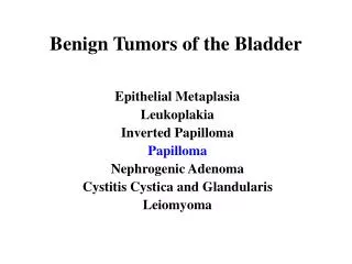
Renal Tumors • Benign: • Oncocytoma • Renal cell adenoma • Malignant: • Renal cell carcinoma • Wilms tumor • Urothelial carcinoma
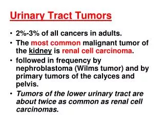
Tumors of the Kidney • Renal cell carcinoma • Arise from tubular epithelium • 85% of primary malignant tumors of the kidney • 2-3% of cancer in adults • 6th-7th decade of life, Men 2x> women • Increased risk in smokers, occupational exposure to cadmium, in dialysis-associated cysts
Clinical: • Hematuria 50% • Pain • Mass • Paraneoplastic syndrome: • Fever, polycythemia 5-10% (erythropoietin) • Hypercalcemia, hypertension, cushing syndrome • Metastases to lung, bone
Renal cell carcinoma: types • 1. Conventional RCC (clear cell) • 2. Papillary RCC • 3. Chromophobe RCC
Renal cell carcinoma: types • 1. Conventional RCC (clear cell RCC) • 70-80% of RCC • Familial and sporadic • Associated with von Hippel-Lindau syndrome • VHL is autosomal dominant • Multiple tumors: hemantioblastoma of cerebellum and retina, renal cysts, renal cell carcinoma • Germline mutation in VHL gene (3p25) • Loss of second allele by somatic mutation • Seen in sporadic RCC as well
Renal cell carcinoma: types • 2. Papillary RCC • 10-15% arise from proximal tubular epithelium • Multifocal, bilateral • Familial and sporadic • MET proto oncogene (7q31) • Trisomy 7, Mutation of chromosome 7 • In sporadic cases: trisomy 7, 16, 17
Renal cell carcinoma: types • 3. Chromophobe RCC: • 5% arise from collecting ducts • Loss of Ch 1,2,6,10,13,17,21 • Hypodiploidy • Good prognosis
Morphology: • Clear cell • Solitary, large, cortical, well defined • Yellow-orange, gray-white, cysts, hemorrhage, necrosis • May extend to pelvis, ureters • May invade renal vein and inferior vena cava • Papillary • Bilateral, multiple • Chromophobe • Brown-tan
Micro: • Clear cell RCC • Lipid, glycogen • Clear cells • Round nuclei • Vascular • Papillary • Chromophobe: • Perinuclear halo, macrovesicles • Well defined cell membrane
Our patient is a 5 year-old, Caucasian female who presented to the primary pediatric clinic in early spring with chief complaints of cough, fever by touch, and decreased activity for six days. Our patient's illness began with rhinorrhea and progressed to appetite loss and fever that her parents felt was unresponsive to acetaminophen.
On physical exam our patient appeared worrisomely “sick”. She was fatigued. The outstanding physical findings consisted of a slightly erythematous throat. On abdominal exam a mass of 9 cm width by 4 cm length with regularly shaped margins was palpated with light depth and verified with percussion in the left upper quadrant. The mass was smooth, slightly firm, oval, nonmobile, and did not cross the midline. The child denied pain during the exam, but was uncomfortable during palpation.
Our patient was then admitted to the children's hospital after her fever and upper respiratory symptoms subsided for biopsy. Biopsy confirmed diagnosis of Wilms' tumor. The tumor was shrunk with chemotherapy for five months and then removed from the left kidney via complete nephrectomy and partial right nephrectomy.
Wilms Tumor (Nephroblastoma) • Most common primary kidney tumor in children • Occur commonly between 2-5 years • WT1 gene, WT2 gene • Risk with congenital malformation: • WAGR syndrome • Denys-Drash syndrome • Beckwith-Weidmann syndorme
Wilms Tumor (Nephroblastoma) • Risk with congenital malformation: • WAGR syndrome • Loss of ch 11p13 (WT1) • Aniridia, genetal abnormalities, mental retardation • Denys-Drash syndrome • Loss of ch 11p13 (WT1) • Gonadal dysgenesis • Renal abnormalities • Beckwith-Weidmann syndrome • Enlarged body organs (tongue, kideny, liver), adrenal enlargement, hemihypertrophy (body segment enlargement) • Ch 11p15.5 (WT2)
Wilms Tumor (Nephroblastoma) • Clinical: • Mass • Cross the midline • Hematuria • Intestinal obstruction • Prognosis: good • 2 year-survival: 90%
Wilms Tumor (Nephroblastoma) • Morphology: • Large well-circumscribed • 10% bilateral, multiple • Soft homogeneous, tan-gray • Hemorrhage, cysts, necrosis • Triphasic: • Epithelial: tubules • Stroma: fibrous, myxoid • Blastema: small blue cells • Foci of anaplasia • Nephtogenic rests: precursor lesions