Download

1 / 35
360 likes | 639 Vues
THE URINARY BLADDER TUMORS D. Diar H. Bajalan. NEOPLASM OF BLADDER. 95 % of primary bladder tumours originate in the epithelium ; The remainder arise from connective tissue ( angioma , myoma , fibroma and sarcoma) .

E N D
THE URINARY BLADDER TUMORS D. Diar H. Bajalan
NEOPLASM OF BLADDER 95 % of primary bladder tumours originate in the epithelium; The remainder arise from connective tissue (angioma, myoma, fibroma and sarcoma) . Secondary tumours: from the sigmoid, rectum, prostate, uterus or ovaries.
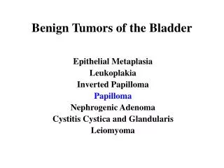
Pathology Benign papillary tumours. The papilloma consists of a single frond of cells with a central vascular core.
It may be of three cell types: 1- Transitional =90%2- Squamous =5%3- Adenocarcinoma =1-2%, which arises usually from the urachal remnant. Carcinoma of the bladder
Transitional cell carcinoma (TCC): • Occupations with an increased risk of bladder cancer: • 1- Textile workers • 2- Dye workers • 3- Tire rubber and cable workers • 4- Petrol workers • 5- Leather workers • 6- Shoe manufacturers and cleaners • 7- Painters • 8- Hairdressers • 9- Lorry drivers • 10- Drill press operators • 11- Chemical workers • 12- Rodent exterminators and sewage workers
Cigarette smoking : is associated with a two to threefold excess risk. In areas where S. haematobiumis endemic bladder cancer is more common, and this tends to be squamous.
Tumour staging and grading Study of the biological behaviour of transitional cell cancer of the bladder shows that they fall into the three following groups: • Non muscle invasive tumours (pTa) and (pT1): account for 70%,may be single or multiple. Histological examination may reveal invasion of the lamina propria (pT1) but not of the muscle or no invasion of lamina propria (pTa) . • Muscle invasive disease(T2)(T3)(T4): accounts for 25%. Such tumours carry a much worse prognosis as they are subject to local invasion and distant metastasis. • Flat, noninvasive carcinoma in situ (CIS) accounts for 5%.
Muscle invasive transitional cell carcinoma • Is nearly always solid. • These tumours are often large and broad based, having an irregular, ugly, sometimes ulcerated appearance. • The incidence of metastases, whether from lymphatic invasion in the pelvis or blood-born to the lung, liver or bones, is much more common and will cause the death of 30—50% of patients.
Pure squamous cell carcinoma of the bladder • It tend to be solid and are nearly always associated with muscle invasion. • This is the most prevalent form of bladder cancer in areas where bilharzia is endemic. • It may be associated with chronic irritation caused by stone disease in the bladder as a result of metaplasia.
Pure ADENOCARCINOMA • 1—2 % of bladder cancer. • It usually arises in the fundus of the bladder at the site of the urachal remnant.
Clinical features • Painless Intermittent haematuria: is by far the most common symptom and should be regarded as indicative of a bladder carcinoma until proven otherwise. • Constant pain in the pelvis usually heralds extravesical spread. • Frequency and dysuria. • Pain in the loin or pyelonephritis may indicate ureteric obstruction and hydronephrosis. • A late manifestation is nerve involvement causing pain referred to the suprapubic region, groins, perineum, anus and into the thighs.
Investigation • Urine:examined cytologically for malignant cells. This is not a good screening test for patients with haematuria as, particularly with low-grade tumours, malignant cells may not be identified . • Blood tests like (Hb , S.electrolytes , B.Urea). • Ultrasound :
IVU: • -The most common radiological sign is a filling defect. • -Hydronephrosis may occur if a superficial tumour grows up into the intramural ureter or if direct invasion of the ureteric wall occurs.
Cystourethroscopy:is the mainstay of diagnosis and should always be performed on patients with haematuria. It can be done with a rigid instrument under general anaesthesia or with a flexible instrument under local anaesthesia. • The urethra is inspected at the initial insertion of the instrument (urethroscopy) and the bladder is then examined in a systematic fashion (cystoscopy). • Bimanual examination. • A bimanual examination with the patient fully relaxed under general anaesthesia should be performed both before and after endoscopic surgical treatment of these tumours.
Noninvasive tumours Endoscopic surgery (TURBT)(Trans Urethral Resection of Bladder Tumor: The tumour should be carefully resected in layers using a resectoscope. The base of the tumour is sent separately for histological examination. Small biopsies are taken near to and distant from the primary lesion to diagnose unsuspected CIS. Follow-up:in patients with a single, low- or medium-grade pTa tumour can safely be treated by resection alone and followed up by means of regular cystoscopies. The resection may be followed by a 6-week course of intravesical chemotherapy with Mitomycin C, Adriamycin or Epirubicin. Others will treat such patients by means of endoscopic treatment followed by intravesical immunotherapy with intravesical bacillus of Calmette and Guérin(BCG). Follow-up cystoscopies are essential:They are usually done in: 3 months intervals in first year. 6 months intervals in the second year. Yearly intervals for the next 3 years.
Intravesical chemotherapy and immunotherapy Various agents have been used as chemotherapy. These include: • Mitomycin C. • Epirubicin. • Adriamycin. • Thiotepa. Immunotherapy agent includes: BCG is now frequently used as intravesical immunotherapy especially in high grades.
Open surgical excision This should be totally avoided. If by some error a bladder containing a tumour is entered, then the tumour may be removed with a diathermy needle and the base coagulated and the bladder closed. Postoperative radiotherapyto the wound will diminish the chance of tumour implantation.
2.Invasive tumours: • Radical cystectomy. • Radical radiotherapy. • Combination of the two. • Primary surgery(radical cystectomy) followed by a combination of agents using Cis-platinum, Methotrexate, Adriamycin and Vinblastine(MVAC)
RADIOTHERAPY • Local radiotherapy: • For small invasive lesions, can be delivered by open placement of a radioactive tantalium wire. It is used infrequently today. • Deep external beam X-ray therapy. • Is usually given by means of high-powered linear accelerators. Radical radiotherapy giving 60 Gy over a 4—6-week period will produce a 40-50% complete response
Surgery • Partial cystectomy: • This should be limited to the treatment of small adenocarcinomas at the dome of the bladder. • Radical cystectomyand pelvic lymphadenectomy. • This is now standard treatment for localised pT2—pT3 disease without evidence of secondary spread. • Needs diversion of urine by: • Ileal conduit. • Ureterosigmodostomy: • Orthotopicilealneobladder
Leukoplakia This condition is simply squamous metaplasia of the bladder. Profuse production of keratin may result in the passing of white particles in the urine. It cannot be treated easily. Localised areas may be resected endoscopically. Diffuse leucoplakia of the bladder is premalignant and results in squamous bladder cancer. Careful cystoscopic assessment is required. The condition may require cystectomy.