Laparoscopic Fundoplication
Gastroesophageal reflux disease (GERD) is defined as the<br>failure of the antireflux barrier, allowing abnormal reflux<br>of gastric contents into the esophagus. It is a condition<br>which develops when the reflux of stomach contents<br>causes troublesome symptoms and complications. It is a<br>mechanical disorder which is caused by a defective lower<br>esophageal sphincter (LES), a gastric-emptying disorder, or<br>failed esophageal peristalsis.


Laparoscopic Fundoplication
E N D
Presentation Transcript
Laparoscopic Fundoplication Prof. Dr. R. K. Mishra INTRODUCTION Symptoms ■ Heartburn (retrosternal burning) ■ Regurgitation ■ Pain ■ Respiratory symptoms Gastroesophageal reflux disease (GERD) is defined as the failure of the antireflux barrier, allowing abnormal reflux of gastric contents into the esophagus. It is a condition which develops when the reflux of stomach contents causes troublesome symptoms and complications. It is a mechanical disorder which is caused by a defective lower esophageal sphincter (LES), a gastric-emptying disorder, or failed esophageal peristalsis. Gastroesophageal reflux is one of the most common digestive symptoms. Exposure of the esophageal mucosa to acid, enzymes, and other digestive secretions leads to acute and chronic inflammations, with pain, and ulceration or stricture formation if untreated. Heartburn occurs in 5–45% of adults in Western countries, depending on the frequency of symptoms, 30–45% suffer from symptoms once a month and 5–10% everyday. The majority of patients suffering from GERD experience minor symptoms for which they do not seek medical attention. Age does not seem to have an impact on the frequency of GERD symptoms, and no causal factor has been identified. Esophagitis due to reflux occurs in approximately 2% of the global population. It is the most frequent form of lesion detected on upper gastrointestinal (GI) endoscopy, occurring more frequently than gastric ulcers or duodenal ulcers. GERD is often a chronic ailment. After a 5–10-year follow-up, about two-thirds of patients complain of persistent symptoms requiring occasional or continuous treatment. Diagnostic Test ■ Endoscopy ■ Barium swallow ■ Esophageal transit +/– manometry ■ pH monitoring Treatment of Gastroesophageal Reflux Disease Medical therapy is the first line of management. Lifestyle modification and medications are the first-line treatment for GERD. Surgical management is generally reserved for patients who have persistent symptoms or develop complications despite optimal medical therapy. Esophagitis will heal in approximately 90% of cases with intensive medical therapy. However, symptoms recur in >80% of cases within 1 year of drug withdrawal. Since, it is a chronic condition, medical therapy involving acid suppression and/ or promotility agents may be required for the rest of patient’s life. Despite the fact that current medical management is very effective for the majority, a small number of patients do not get complete relief of symptoms. Currently, there is increasing interest in the surgical management of GERD. The goal of surgical therapy is to recreate an antireflux barrier. Surgical management of GERD focuses on restoring a physiologic equivalent to the normal LES. It is the only treatment capable of changing the natural history of GERD. This interest in surgical therapy has been renewed with the advent of laparoscopic surgery. PATHOPHYSIOLOGY The pathophysiology of GERD is multifactorial, although it is usually due to the weakening of the anatomical or functional gastroesophageal barrier located at the esophagogastric junction. Injury to the esophageal mucosa by acid-peptic gastric secretions, while secondary to this weakening, plays a major role in the development of GERD symptoms and lesions. In fact, suppressing the gastric acid secretion, which is the usual treatment of this ailment, leads to the disappearance of symptoms and healing of lesions in almost all cases. GERD is, therefore, acid-dependent. Indications for Surgical Treatment Currently, there is increasing interest in the surgical management of GERD. There are a number of reasons for this. Despite the fact that current medical management is very effective for the majority, a small number of patients
270 SECTION2: Laparoscopic General Surgical Procedures Upper Gastrointestinal Endoscopy Esophageal and gastric endoscopy should be performed to assess the esophageal and gastric mucosa for signs of malignancy prior to proceeding with an antireflux procedure. Patients with endoscopic findings of severe (Los Angeles grade C or D) esophagitis, biopsy-proven Barrett’s esophagus ≥1 cm, or a benign peptic stricture are good candidates for fundoplication surgery. do not get complete relief of symptoms. Secondly, some patients, particularly those who are in their 20s or 30s, face the prospect of a lifetime of continuous proton- pump inhibitor (PPI) therapy with the possible risk of, as yet, unknown side effects. In addition, the laparoscopic approach with its benefits of reduced operative trauma and less time off work has become a more common place. As a consequence, general practitioners and gastroenterologists are more convinced to refer patients with disabling symptoms for surgical treatment. Antireflux surgery is most often performed to control GI symptoms such as heartburn and regurgitation that are refractory to medical therapy. It may also be performed for non-GI symptoms such as chronic cough, laryngeal disease, and asthma. If there is solid objective evidence to attribute such symptoms to reflux. The GI symptoms are also referred to as typical symptoms and the non-GI symptoms as atypical symptoms. The gold standard antireflux operation is undoubtedly the Nissen type of total fundoplication and many studies have affirmed its effectiveness in controlling acid reflux. However, new symptoms after fundoplication such as gas bloat and dysphagia, which probably result from a hypercompetent LES produced by the Nissen operation, are common. Concerning the indication for surgery, a distinction between heartburn and regurgitation symptoms is considered an important factor (medical treatment appears to be more effective for heartburn than for regurgitation). Even after successful medical acid suppression, the patient can have recurrent symptoms of epigastric pain and retrosternal pressure as well as food regurgitation due to an incompetent cardia, insufficient peristalsis, or a large hiatal hernia. Surgical therapy should be considered in individuals with documented GERD who are: ■ Refractory to medical management ■ Associated with hiatus hernia ■ Intolerance to postpartum hemorrhage (PPH) or H2- receptors ■ Not compliant to medical therapy ■ Have complications of GERD, e.g., Barrett’s esophagus, stricture, and grade 3 or 4 esophagitis ■ Atypical symptoms such as asthma, hoarseness, cough, chest pain, and aspiration Study has shown those patients resistant to anti secretory treatment are not good candidates for antireflux surgery. Esophageal pH Testing Esophageal pH monitoring is the gold standard technique for the detection of gastroesophageal reflux in adults and children. Patients with typical symptoms of GERD should undergo standard pH testing. Patients with atypical symptoms may require nonstandard pH testing, such as one with dual pH probe or impedance in order to document proximal and/or nonacidic reflux. Standard pH Testing Ambulatory pH testing is the gold standard for diagnosing pathologic GERD. Prior to antireflux surgery, all patients with nonerosive GERD, including those with Los Angeles grade A or B, esophagitis, and those with short-segment (<1 cm) Barrett’s esophagus, should undergo standard pH testing to document abnormal distal esophageal acid exposure. An abnormal pH test in a PPI-dependent patient with typical symptoms predicts successful outcomes with antireflux surgery. Standard pH testing can be done via a transnasal catheter for 24 hours or a wireless pH system for 48 hours after the patient has been off acid suppression for 7 days. Multichannel Intraluminal Impedance Testing Multichannel intraluminal impedance (MII) is a catheter- based method to detect intraluminal bolus movement within the esophagus. MII is performed in combination with manometry or pH testing. Combined MII and pH (MII-pH) testing can detect both acid and nonacid gastroesophageal reflux. It should be recognized that this testing is somewhat controversial and interpretation is operator-dependent. Dual pH Probe Despite other available and seemingly superior technologies such as impedance testing, the current “gold standard” diagnosis of extraesophageal reflux continues to be 24-hour dual pH probe monitoring. This procedure involves placing a catheter through the nose and into the esophagus. Two sensors, proximally and distally located within the catheter, detect the pH level or acidity in the distal esophagus and hypopharynx. Extraesophageal reflux is diagnosed when stomach contents are shown to flow upward from the stomach PREOPERATIVE EVALUATION All the patients should have endoscopy, standard pH testing, esophageal manometry, and barium esophagram be performed before antireflux surgery. Nonstandard pH testing or gastric-emptying study may be required for some patients.
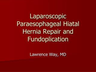
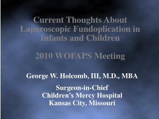
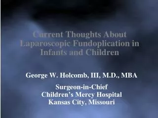
271 CHAPTER20: Laparoscopic Fundoplication to the distal esophagus, and subsequently to the proximal esophagus and into the hypopharynx. Measurements of pH are recorded to a small, portable device that is worn on a belt during the study. to pass into the stomach, but prevent stomach acid from flowing into the esophagus and thus prevent GERD. Laparoscopic fundoplication is a useful method for reducing hospital stay, complications, and early return to normal activity. Esophageal Manometry Esophageal manometry is the most reliable way to assess LES competence and esophageal peristalsis. Overall, the LES is incompetent in about 60% of patients with GERD, while transient relaxation of a competent LES is the cause of GERD in the remaining 40%. Manometry assesses esophageal peristalsis as well and occasionally provides alternative diagnoses, such as scleroderma or achalasia, for which antireflux surgery may be contraindicated. Manometric findings may influence the surgical approach such as partial instead of complete fundoplication for those with weak peristalsis. TYPES OF FUNDOPLICATION SURGERY Laparoscopic fundoplication has become the standard surgical method of treating GERD. Although Nissen total fundoplication is the most commonly performed procedure, partial fundoplication, either anterior or posterior, is becoming more acceptable because of a suggested lower risk of long-term side effects (Fig. 1). The 360° Nissen fundoplication has been the standard operation for gastroesophageal reflux, but is associated with substantial rates of “gas bloat,” gagging, and dysphasia (Fig. 2). Toupet fundoplication (TF), a 270° posterior wrap, has fewer complications, and its outcome in compared with Nissen fundoplication, is favorable both in children as well as adults (Fig. 3). Although Nissen total fundoplication is the most commonly performed procedure, partial fundoplication, either anterior or posterior, is becoming more acceptable because of lower risk of long-term complication. Dor in 1962 described anterior fundoplication as an antireflux operation for patients who had a Heller myotomy for achalasia. In the 1970s, Watson developed an operation for patient suffering from GERD. Most surgeons believe that the TF, a 270° posterior wrap, originally described in conjunction with myotomy for achalasia, has fewer complications, and its long-term outcome in comparison with NF is favorable both in children as well as adults. The main tasks of this operation consist of: ■ Preparation of the patient ■ Creation of pneumoperitoneum and insertion of ports ■ Diagnostic laparoscopy and dissection of visceral peritoneum Barium Esophagram Barium esophagram can demonstrate esophageal length, presence, and size of any hiatal hernia, presence of any esophageal diverticulum or stricture, and the extent of reflux with provocation. Gastric-emptying Study A 4-hour gastric-emptying study should be performed when the history of a patient with GERD symptoms suggests gastric outlet obstruction or gastroparesis with significant nausea, repeated vomiting, severe bloating or postprandial fullness, or retained food in the stomach after overnight fast. METHODS OF FUNDOPLICATION There is no one best operation for all patients with GERD. Currently available antireflux procedures include: ■ Endoscopic radiofrequency treatment (Stretta) ■ Endoscopic transoral incisionless fundoplication (TIF) ■ Laparoscopic magnetic sphincter augmentation (LINX) ■ Laparoscopic Hill gastropexy ■ Laparoscopic partial fundoplication ■ Laparoscopic Nissen fundoplication (NF) Laparoscopic fundoplication is a safe procedure and can provide less postoperative morbidity in experienced hands. Fundus of the stomach, which is on the left of the esophagus and main portion of the stomach, is wrapped around the back of the esophagus until it is once again in front of this structure. The portion of the fundus that is now on the right side of the esophagus is sutured to the portion on the left side to keep the wrap in place. The fundoplication resembles a buttoned shirt collar. The collar is the fundus wrap and the neck represents the esophagus imbricated into the wrap. This has the effect of creating a one-way valve in the esophagus to allow food Fig. 1: Selection of type of fundoplication. (GERD: gastroesophageal reflux disease; LES: lower esophageal sphincter)
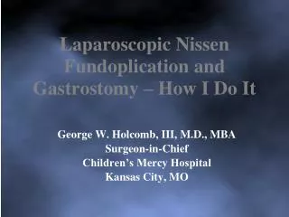
272 SECTION2: Laparoscopic General Surgical Procedures Fig. 2: Nissen fundoplication. Fig. 3: Toupet fundoplication (TF), a 270° wrap. ■ ■ ■ Mobilization of 5 cm of intra-abdominal esophagus Fundus pull from below the esophagus Insertion of posterior sutures to tighten the crural opening Fixation of fundus to the left crura Fixation of the fundus with the right crura Fixation of the fundus with esophagus. Inspection of tightness of fundoplication Irrigation and suction of operating field Final diagnostic laparoscopy for any bowel injury or hemorrhage Removal of the instrument with complete exit of CO2. Closure of wound. ■ ■ ■ ■ ■ ■ Fig. 4: Laparoscopic fundoplication port position. PATIENT SELECTION Patient Position Many patients have symptoms palliated by lifestyle such as diet and exercise, others by simple medication, and some by strong medication such as PPI. A certain proportion of patients have refractory or long-term symptoms, and operation can be considered in this group of patients. As reflux symptoms are frequent and variable, it is wise to obtain both ambulatory 24 hours pH-metry and esophageal motility studies prior to surgery. Upper GI endoscopies should be performed in all patients. The patient is placed on the operating table with the legs in stirrups, the knees slightly bent, and the hips flexed approximately 10°. The operating table is tilted head up by approximately 15°. Compression bandage is used on leg during the operation to prevent thromboembolism. The surgeon stands between the patient’s legs. The first assistant, whose main task is to position the video camera, sits on the surgeon’s left side. The instrument trolley is placed on the patient’s left allowing the scrub nurse to assist with placing the appropriate instruments in the operating ports. Television monitors are positioned on either side of the top end of the operating table at a suitable height for surgeon, anesthetist, and assistant to see the procedure. OPERATIVE TECHNIQUE The original fundoplication as described by Rudolph Nissen in 1956 involved passage of the gastric fundus behind the esophagus to encircle the distal 5 cm of the esophagus. In contemporary surgical practice, fundoplication is performed laparoscopically in most patients. Compared with open transabdominal or transthoracic fundoplication, laparoscopic fundoplication demonstrates comparable safety, efficacy in symptom relief, and patient satisfaction. Port Position The 10-mm camera (port 1) is placed in the midline approximately 5 cm above the umbilicus; this position will vary depending on the build of the patient (Fig. 4). After
273 CHAPTER20: Laparoscopic Fundoplication inserting the camera, a 5-mm port (2) is inserted in the right upper quadrant 8–10 cm from the midline. A port (3), with a variable 5–10 mm size, is placed in the left upper quadrant—a mirror image of the one on the patient’s right. This allows both 5 mm and 10 mm instruments to be used through the same cannula without changing ports. A further 5-mm port (4) is positioned in the left anterior axillary line immediately below the costal margin. This port is mainly used for a forceps which will hold the tape encircling the esophagus (Fig. 5). Liver retraction used to be one of the more problematic aspects of laparoscopic fundoplication. In our experience, these difficulties have been largely overcome by the use of the Nathanson liver retractor. Alternate port position in laparoscopic fundoplication is shown in Figures 6A and B. surrounding loose areolar tissues. The loose areolar tissue around the esophagus is exposed and secures the bleeding from any blood vessels visible during mobilization of esophagus. Always remember not to injure the esophageal wall and vagal fibers when dissecting this area around esophagus. The space between the hiatus and the anterior aspect of esophagus is developed using fine dissection by scissors to divide blood vessels crossing this space. Lesser Sac Opened The posterior aspect of the left crus is identified as it meets the right crus and dissection of its surface commences, especially the peritoneal covering over the margin of right crus is dissected down to the fundus from the diaphragm known as Rossetti dissection technique. Dissection of the posterior aspect of the left crus is done by lifting the intra- abdominal esophagus forward with a blunt instrument. A sling is fed into the jaws of the grasping forceps and then pulled round behind the esophagus (Fig. 8). Tissue Dissection and Mobilization Mobilization of Stomach The fundus part of the stomach is mobilized nicely along with the gastroesophageal junction by anteromedial traction. The short gastric vessels are separated with the help of a bipolar or harmonic scalpel which aids in further mobilization of the fundus of the stomach through the earlier formed posterior window. Care must be taken not to injure the spleen which is adjacent to the stomach. Mobilization of Esophagus Dissection starts at the avascular portion of the lesser omentum above the hepatic branch of the vagus. The dissection is continued carefully up to the hiatus, which can be seen through the defect created. An opening is created in the lesser omentum, above the hepatic branch of vagus to allow better access to the hiatus (Figs. 7A to H). The right crus is dissected using electrosurgery and scissors to identify the plane between the crus and Fig. 5: Port position in fundoplication. A B Figs. 6A and B: Alternative port position in laparoscopic fundoplication.
274 SECTION2: Laparoscopic General Surgical Procedures Sling is passed through a separate punctured wound from abdominal wall without port. A grasping forceps is inserted through one of the port to hold the sling so that the esophagus can be manipulated. Dissection surroundings of esophagus in the posterior mediastinum should for approximately 5–6 cm. Mobilization of the esophagus and stomach should be sufficient enough to have a good floppy fundus for wrap (Fig. 9). The next step is to pull the fundus from behind the esophagus to form a wrap. A B C D E F G H Figs. 7A to H: Mobilization of esophagus.
275 CHAPTER20: Laparoscopic Fundoplication Fundus Pull After mobilizing the fundus nicely, the tip of the fundus is pulled by one of the grasper introduced through below and right side of the esophagus (Figs. 10 and 11). The mobilization of stomach should be adequate to give a floppy fundus for plication otherwise patient may develop dysphagia. One stay suture may be applied to the fundus to hold it in place or one of the grasper may be used to keep it pulled. The next step is crural repair. is made around the esophagus, this can be ascertained by seeing the “buttock sign.” The wrap made by both posterior and anterior walls of fundus should not be too tight. A Floppy Nissen’s is made by doing the “shoe shine” maneuver. Suture material used is nonabsorbable sutures such as Dacron, silk, or polypropylene. Approximately 5 cm of mobilized esophagus should be prepared for fundal wrap. First suture is taken with full thickness of anterior wall of fundus, and full thickness of posterior wall of fundus is taken. Intracorporeal surgeon’s knot are used for plicating the stomach on to the esophagus, though “tumble square knots” can also be used. Two sutures are taken above the first knot at a distance of 1 cm between each knot, involving full thickness of anterior wall of fundus, partial muscular layer of esophagus, and full thickness posterior wall of fundus. In the above suture, wall of esophagus is sandwiched between walls of fundus. Taking full thickness of esophagus in above sutures should be avoided as it may cause a complication of perforation of Crural Approximation A posterior crural repair should be performed by approximating the right and left pillar of the left crus with sutures. Crura should be approximated behind the esophagus using two or three sutures of 2/0 braided polyamide on a 30-mm needle using tumble square knot or surgeon’s knot. A further one or two sutures are inserted in the same way, at about 1 cm intervals and tied. It is important not to make the crural opening too tight since this will produce dysphagia (Figs. 12A to D). Further three sutures are then positioned at approximately 1 cm intervals to the posterior fundus and the right crus. The sling used for esophageal retraction is removed. A 50–60 French bougie should be placed in the stomach along the lesser curvature to calibrate the tightness of the fundoplication. Fundoplication By using an atraumatic grasper in the left hand, the posterior wall of the fundus is held through the opening made behind the esophagus. The posterior wall of fundus is pulled gently and at the same time, the extra-anterior wall is pushed back to avoid crowding of tissue within the window. By serial movements of pulling posterior wall forward and pushing anterior wall back, a sufficient amount of wrap Fig. 9: Sling application around esophagus for proper exposure of crura. Fig. 8: Sling insertion. Fig. 10: Mobilized fundus of stomach is pulled from behind the esophagus.
276 SECTION2: Laparoscopic General Surgical Procedures A B Figs. 11A and B: Fundus of stomach is pulled from behind the esophagus. A B C D Figs. 12A to D: Approximation of crura. esophagus. Two sutures are taken below the first knot at a distance of 1 cm between each knot in similar manner. Most surgeons would perform a wrap of 2–3 cm (Figs. 13A to C). should be removed carefully. Remove telescope leaving gas valve of umbilical port open to let out all the gas. Close the wound with suture. Use vicryl for rectus and unabsorbable intradermal or stapler for skin. Apply adhesive sterile dressing over the wound. Patient may be discharged 2 days after operation, if everything goes well. Long-term observational studies of Ending of the Operation Abdomen should be examined for any possible bowel injury or hemorrhage. The instrument and then port
277 CHAPTER20: Laparoscopic Fundoplication A B C Figs. 13A to C: Fixation of wrap by intracorporeal sutures. laparoscopic fundoplication performed by experienced operators generally report that 90–95% of adult patients are satisfied with the results of their surgery. The patient may have slight dysphagia initially but usually resolves after 6 weeks. The patient having any complain of dysphagia should be examined endoscopically after 3–4 weeks of operation. Dysphagia that persists for >12 weeks requires evaluation, which typically begins with a barium swallow to assess the anatomic placement of the fundoplication. In case of tight plication endoscopic dilatation technique, bougie or guidewire dilation should be done. Endoscopic dilatation procedure is well tolerated and produces good results. Patients who have a 360° fundoplication may be candidates for revision to a partial fundoplication, if dysphagia persists and effective barium tablet passage cannot be established despite dilation. Symptoms of gas bloat syndrome can be elicited in a significant number of patients after fundoplication. Symptoms tend to lessen over time in most patients. In patients with severe persistent symptoms despite the medical treatment approaches, pyloroplasty, pyloric Botox injection, and pneumatic pyloric dilatation are options in select patients who have documented gastroparesis. Long-term mortality of laparoscopic fundoplications is very low. The reported 30-day surgical mortality rate of laparoscopic fundoplication is <0.1–0.2%. The most commonly reported acute complications for laparoscopic fundoplications are gastric or esophageal injury, splenic injury or splenectomy, pneumothorax, bleeding, pneumonia, fever, wound infections, bloating, and dysphagia. Major acute complications are uncommon. In a study of 2,655 patients, 4.1% had a complication within 30 days of surgery, including 1.1% infection, 0.9% bleeding, and 0.9% esophageal perforation. Rossetti–Nissen Fundoplication A common modification is a 360° fundic wrap without division of the short gastric vessels is called Rossetti–Nissen fundoplication. However, most surgeons prefer to divide the short gastric vessels to allow for greater freedom of mobilization and reduced concern about torque on the fundoplication which is hard to measure and also facilitate lower esophageal relaxation. Partial Fundoplications A partial 270° posterior wrap, TF is used for patients with severe associated motor abnormalities. A partial 180° anterior wrap (Dor) has also been described. Based on currently available data, the choice of anterior versus
278 SECTION2: Laparoscopic General Surgical Procedures for GERD (Figs. 14A and B). The device works by augmenting the LES with a ring made up of a series of rare earth magnets. The magnets have sufficient attraction to increase the LES closure pressure but permit food passage with swallowing. Eligible patients must demonstrate typical GERD symptoms, an abnormal pH study, partial response to daily PPI therapy, and absence of a large hiatal hernia (>3 cm) or severe esophagitis. Patients who have allergies to titanium, stainless steel, nickel, or iron should not receive a LINX prosthesis. Patients who have the LINX device need to consult their surgeon before undergoing magnetic resonance imaging (MRI). A new version of LINX considered “MR Conditional” in a MRI system up to 1.5 Tesla (1.5 T) is now available. Scanning under different conditions may interfere with the magnetic strength and function of the device. Laparoscopic magnetic sphincter augmentation device has been compared with laparoscopic Nissen fundo- plication in several meta-analyses. Clinical control of reflux was comparable after the two procedures in both the short and longer term. In the longer term, there was less gas bloat associated with LINX than fundoplication. Presently, however, there is insufficient evidence that would allow surgeons to choose between the two procedures. posterior or partial versus complete NF should be left to the individual surgeon. Hill Gastropexy The Hill procedure involves imbrication of the anterior and posterior collar sling muscular fibers at the level of the gastroesophageal junction around the esophagus with tethering of the complex to the median arcuate ligament and closure of the diaphragm. Intraoperative manometry is used to achieve a desired LES pressure. This operation can also be used in a patient with a small stomach because of prior gastric resection. In a randomized trial, laparoscopic Hill gastropexy and laparoscopic Nissen fundoplication yielded similar and both excellent outcomes at 1-year follow-up. In a single-center retrospective study with a median follow-up of 18.5 years, 85% of patients who underwent Hill gastropexy reported good to excellent symptomatic outcomes and quality of life. Magnetic Sphincter Augmentation (LINX Device) In 2012, the US Food and Drug Administration (FDA) approved the LINX reflux management system as a treatment A B Figs. 14A and B: LINX sphincter augmentation system. Fig. 15: Stretta therapy as a treatment option for gastroesophageal reflux disease (GERD).
279 CHAPTER20: Laparoscopic Fundoplication A B D E C Figs. 16A to E: Transoral incisionless fundoplication (TIF). A B Figs. 17A and B: EsophyX device for transoral incisionless fundoplication (TIF).
280 SECTION2: Laparoscopic General Surgical Procedures Stretta Endoscopic Procedure The Stretta procedure is the most widely studied endoscopic antireflux procedure. The specialized catheter is placed with endoscopic assistance over a guidewire. Using monopolar energy, a series of 56 treatments is delivered across five levels. Stretta is rated as effective therapy in patients with a lower esophageal pressure of at least 8 mm Hg and hiatal hernia <3 cm. The energy heats the tissue, ultimately causing it to swell and stiffen; the way this works was not understood as of 2019, but it was thought that perhaps the heat causes local inflammation, collagen deposition, and muscular thickening of the LES and that it may disrupt the nerves there (Fig. 15). BIBLIOGRAPHY 1. Akinola E, Rosenkrantz TS, Pappagallo M, McKay K, Hussain N. Gastroesophageal reflux in infants <32 weeks gestational age at birth: lack of relationship to chronic lung disease. Am J Perinatol. 2004;21(2):57-62. 2. Anvari M, Bamehriz F. Outcome of laparoscopic Nissen fundoplication in patients with body mass index >or= 35. Surg Endosc. 2006;20(2):230-4. 3. Augood C, MacLennan S, Gilbert R, Logan S. Cisapride treatment for gastroesophageal reflux in children. Cochrane Database Syst Rev. 2003;(4):CD002300. 4. Boyce PM, Talley NJ, Burke C, Koloski NA. Epidemiology of the functional gastrointestinal disorders diagnosed according to Rome II criteria: an Australian population-based study. Int Med J. 2006;36(1):28-36. 5. Bredenoord AJ, Weusten BL, Smout AJ. Symptom association analysis in ambulatory gastroesophageal reflux monitoring. Gut. 2005;54(12):1810-7. 6. Cameron BH, Blair GK, Murphy JJ III, Fraser GC. Morbidity in neurologically impaired children after percutaneous endoscopic versus Stamm gastrostomy. Gastrointest Endosc. 1995;42(1):41-4. 7. Cezard JP. Managing gastroesophageal reflux disease in children. Digestion. 2004;69(1):3-8. 8. Chicella MF, Batres LA, Heesters MS, Dice JE. Prokinetic drug therapy in children: a review of current options. Ann Pharmacother. 2005;39(4):706-11. 9. Chitkara DK, Fortunato C, Nurko S. Esophageal motor activity in children with gastroesophageal reflux disease and esophagitis. J Pediatr Gastroenterol Nutr. 2005;40(1):70-5. 10. Chung DH, Georgeson KE. Fundoplication and gastrostomy. Semin Pediatr Surg. 1998;7(4):213-9. 11. Colletti RB, Di Lorenzo C. Overview of pediatric gastro esophageal reflux disease and proton pump inhibitor therapy. J Pediatr Gastroenterol Nutr. 2003;37(1):S7-S11. 12. Corey KE, Schmitz SM, Shaheen NJ. Does a surgical antireflux procedure decrease the incidence of esophageal adenocarcinoma in Barrett's esophagus? A meta-analysis. Am J Gastroenterol. 2003;98(11):2390-4. 13. Costantini M, Crookes PF, Bremner RM, Hoeft SF, Ehsan A, Peters JH, et al. Value of physiologic assessment of foregut symptoms in a surgical practice. Surgery. 1993;114(4):780-6. 14. Craig WR, Hanlon-Dearman A, Sinclair C, Taback S, Moffatt M. Metoclopramide, thickened feedings, and positioning for gastroesophageal reflux in children under two years. Cochrane Database Syst Rev. 2004;(4):CD003502. 15. Curry JI, Lander TD, Stringer MD. Review article: erythromycin as a prokinetic agent in infants and children. Aliment Pharmacol Ther. 2001;15(5):595-603. 16. D’Alessio MJ, Arnaoutakis D, Giarelli N, Villadolid DV, Rosemurgy AS. Obesity is not a contraindication to laparo scopic fundoplication. J Gastrointest Surg. 2005;9(7):949-54. 17. Davidson GP, Omari TI. Pathophysiological mechanisms of gastroesophageal reflux disease in children. Curr Gastroenterol Rep. 2001;3(3):257-62. 18. Downing TE, Sporn TA, Bollinger RR, et al. Pulmonary histopathology in an experimental model of chronic aspiration is independent of acidity. Exp Biol Med (Maywood). 2008;233(10):1202-12. 19. El-Serag HB, Sweet S, Winchester CC, Dent J. Update on the epidemiology of gastro-oesophageal reflux disease: a systematic review. Gut. 2014;63(6):871-80. 20. Esposito C, Langer JC, Schaarschmidt K, Mattioli G, Sauer C, Centonze A, et al. Laparoscopic antireflux procedures in the management of gastroesophageal reflux following esophageal atresia repair. J Pediatr Gastroenterol Nutr. 2005;40(3):349-51. Transoral Incisionless Fundoplication Transoral incisionless fundoplication is an endoscopic procedure performed under general anesthesia to create a full-thickness serosa-to-serosa plication (Figs. 16A to E). TIF creates 3–5 cm in length and 200–300° in circumference partial fundoplication. TIF can be performed in patients with typical GERD symptoms, no or only low-grade erosive esophagitis (grades A and B), and no or only small hiatal hernia (≤2 cm). TIF is contraindicated in patients with high- grade erosive esophagitis, Barrett’s esophagus, atypical and extraesophageal symptoms of GERD, scleroderma, or other esophageal pathology or surgery. The EsophyX device has been CE marked for the market and FDA approved for the United States market for TIF procedures (Figs. 17A and B). Transoral incisionless fundoplication has been compared with PPI therapy in several randomized trials. In the latest trial (TEMPO), 63 patients with GERD refractory to PPI received TIF versus maximum standard dose PPI therapy. At 6 months, both regurgitation and extraesophageal symptoms were eliminated in more TIF than PPI patients (62 vs. 5%); 90% of TIF patients were off PPIs. After 6 months, all patients in the PPI (control) group elected to cross-over to TIF. At 3 years, 90% and 88% of patients reported elimination of troublesome regurgitation and all atypical symptoms, respectively. Although TIF is associated with fewer postoperative adverse effects such as gas bloating and dysphagia compared with surgical fundoplication, the durability of TIF is less certain than that of surgery. For most patients undergoing surgical treatment for GERD, laparoscopic NF is a better option. This procedure appears to be the most effective and durable amongst all the procedures but is also associated with the greatest potential for dysphagia, difficulty in vomiting, and gas bloating. Patients with decreased esophageal motility or morbid obesity may benefit from an alternative procedure. For an individual patient, the best procedure may be the procedure in which the surgeon is most skilled.
281 CHAPTER20: Laparoscopic Fundoplication 21. Eubanks TR, Omelanczuk P, Richards C, Pohl D, Pellegrini CA. Outcomes of laparoscopic antireflux procedures. Am J Surg. 2000;179(5):391-5. 22. Farrell TM, Richardson WS, Halkar R, Lyon CP, Galloway KD, Waring JP, et al. Nissen fundoplication improves gastric motility in patients with delayed gastric emptying. Surg Endosc. 2001;15(3):271-4. 23. Franco MT, Salvia G, Terrin G, Spadaro R, De Rosa I, Iula VD, et al. Lansoprazole in the treatment of gastroesophageal reflux disease in childhood. Dig Liver Dis. 2000;32(8):660-6. 24. Fraser J, Watson DI, O’Boyle CJ, Jamieson GG. Obesity and its effect on outcome of laparoscopic Nissen fundoplication. Dis Esophagus. 2001;14(1):50-3. 25. Galvani C, Fisichella PM, Gorodner MV, Perretta S, Patti MG. Symptoms are a poor indicator of reflux status after fundoplication for gastroesophageal reflux disease. Arch Surg. 2003;138(5):514-9. 26. Georgeson K. Personal experience with laparoscopic fundoplication in children. Birmingham, AL: Elsevier; 2005. 27. Georgeson KE. Laparoscopic fundoplication and gastrostomy. Semin Laparosc Surg. 1998;5(1):25-30. 28. Gilger MA, Yeh C, Chiang J, Dietrich C, Brandt ML, El-Serag HB. Outcomes of surgical fundoplication in children. Clin Gastroenterol Hepatol. 2004;2(11):978-84. 29. Gold BD. Outcomes of pediatric gastroesophageal reflux disease: in the first year of life, in childhood, and in adults …oh, and should we really leave Helicobacter pylori alone? J Pediatr Gastroenterol Nutr. 2003;37(1):S33-9. 30. Gregersen H, Drewes AM. Functional findings in irritable bowel syndrome. World J Gastroenterol. 2006;12(18):2830-8. 31. Gwee K, Chua ASB. Functional dyspepsia and irritable bowel syndrome, are they different entities and does it matter? World J Gastroenterol. 2006;12(17):2708-12. 32. Hassall E. Decisions in diagnosing and managing chronic gastroesophageal reflux disease in children. J Pediatr. 2005;146(3 Suppl):S3-12. 33. Hassall E. Wrap session: Is the Nissen slipping? Can medical treatment replace surgery for severe gastroesophageal reflux disease in children? Am J Gastroenterol. 1995;90(8):1212-20. 34. Hatch KF, Daily MF, Christensen BJ, Glasgow RE. Failed fundoplications. Am J Surg. 2004;188(6):786-91. 35. Jamieson GG, Duranceau A. What is a Nissen fundoplication? Surgery. 1984;159(6):591-3. 36. Jamieson JR, Stein HJ, DeMeester TR, Bonavina L, Schwizer W, Hinder RA, et al. Ambulatory 24-h esophageal pH monitoring: normal values, optimal thresholds, specificity, sensitivity, and reproducibility. Am J Gastroenterol. 1992;87(9):1102-11. 37. Jesch NK, Schmidt AI, Strassburg A, Gluer S, UreBM. Laparoscopic fundoplication in neurologically impaired children with percutaneous endoscopic gastrostomy. Eur J Pediatr Surg. 2004;14(2):89-92. 38. Johanson JF. Epidemiology of esophageal and supraesophageal reflux injuries. Am J Med. 2000;108 Suppl 4a:99S. 39. Jones R, Canal DF, Inman MM, Rescorla FJ. Laparoscopic fundoplication: a three-year review. Am Surg. 1996;62(8):632-6. 40. Katz PO, Gerson LB, Vela MF. Guidelines for the diagnosis and management of gastroesophageal reflux disease. Am J Gastroenterol. 2013;108(3):308-28. 41. Khajanchee YS, O’Rourke RW, Lockhart B, Patterson EJ, Hansen PD, Swanstrom LL. Postoperative symptoms and failure after antireflux surgery. Arch Surg. 2002;137(9):1008-14. 42. Klauser AG, Schindlbeck NE, Muller-Lissner SA. Symptoms in gastroesophageal reflux disease. Lancet. 1990;335(8683):205-8. 43. Langer JC. The failed fundoplication. Semin Pediatr Surg. 2003;12(2):110-17. 44. Lazarchik DA, Frazier KB. Dental erosion and acid reflux disease: an overview. Gen Dent. 2009;57(2):151-6. 45. Lindquist SG, Kirchhoff M, Lundsteen C, Pedersen W, Erichsen G, Kristensen K, et al. Further delineation of the 22q13 deletion syndrome. Clin Dysmorphol. 2005;14(2):55-60. 46. Liu DC, Somme S, Mavrelis PG, Hurwich D, Statter MB, Teitelbaum DH, et al. Stretta as the initial antireflux procedure in children. J Pediatr Surg. 2005;40(1):148-51; discussion 151-42. 47. Lord RVN, Kaminski A, Oberg S, Bowrey DJ, Hagen JA, DeMeester SR, et al. Absence of gastroesophageal reflux disease in a majority of patients taking acid suppression medications after Nissen fundoplication. J Gastrointest Surg. 2002;6(1):3-9. 48. Ludemann R, Watson DI, Jamieson GG, Game PA, Devitt PG. Five-year follow-up of a randomized clinical trial of laparoscopic total versus anterior 180° fundoplication. Br J Surgery. 2005;92(2):240-3. 49. Madan A, Minocha A. Despite high satisfaction, majority of gastroesophageal reflux disease patients continue to use proton pump inhibitors after antireflux surgery. Aliment Pharmacol Ther. 2006;23(5):601-5. 50. Maddern GJ. The reproducibility of esophageal manometry. Dis Esophagus. 1991;4(2):95-9. 51. Maret-Ouda J, Konings P, Lagergren J, Brusselaers N. Antireflux surgery and risk of esophageal adenocarcinoma: a systematic review and meta-analysis. Ann Surg. 2016;263(2):251-7. 52. Mattioli G, Sacco O, Repetto P, Pini Prato A, Castagnetti M, Carlini C, et al. Necessity for surgery in children with gastroesophageal reflux and supraesophageal symptoms. Eur J Pediatr Surg. 2004;14(1):7-13. 53. Ollyo JB, Lang F, Fontolliet CH, Monnier P. Savary-Miller’s new endoscopic grading of reflux esophagitis: a simple, reproducible, logical, complete and useful classification. Gastroenterology. 1990;89:A100. 54. Papasavas PK, Keenan RJ, Yeaney WW, Caushaj PF, Gagne DJ, Landreneau RJ. Effectiveness of laparoscopic fundoplication in relieving the symptoms of gastroesophageal reflux disease (GERD) and eliminating antireflux medical therapy. Surg Endosc. 2003;17(8):1200-5. 55. Patterson EJ, Davis DG, Khajanchee Y, Swanstrom LL. Comparison of objective outcomes following laparoscopic Nissen fundoplication versus laparoscopic gastric bypass in the morbidly obese with heartburn. Surg Endosc. 2003;17(10):1561-5. 56. Patti MG. An evidence-based approach to the treatment of gastroesophageal reflux disease. JAMA Surg. 2016;151(1):73-8. 57. Post JC, Ze F, Ehrlich GD. Genetics of pediatric gastroesophageal reflux. Curr Opin Allergy Clin Immunol. 2005;5(1):5-9. 58. Quigley EMM. Changing face of irritable bowel syndrome. World J Gastroenterol. 2006;12(1):1-5. 59. Ravelli AM, Milla PJ. Vomiting and gastroesophageal motor activity in children with disorders of the central nervous system. J Pediatr Gastroenterol Nutr. 1998;26(1):56-63. 60. Rothenberg SS. Laproscopic redo-Nissen fundoplication in infants and children. Surg Endosc. 2006;20(10):1518-20. 61. Rothenberg SS. The first decade’s experience with laparoscopic Nissen fundoplication in infants and children. J Pediatr Surg. 2005;40(1):142-6; discussion 147. 62. Rudolph CD. Supraesophageal complications of gastro esophageal reflux in children: challenges in diagnosis and treatment. Am J Med. 2003;115(Suppl 3A):150S-6S. 63. Sages (2010). Guidelines for surgical treatment of gastroesophageal reflux disease (GERD). Society of American Gastrointestinal and Endoscopic Surgeons (SAGES). [online] Available from: https:// www.sages.org/publications/guidelines/guidelines-for-surgical- treatment-of-gastroesophageal-reflux-disease-gerd/. [Last accessed August, 2020). 64. Schier F. Indications for laparoscopic antireflux procedures in children. Semin Laparosc Surg. 2002;9(3):139-45.
282 SECTION2: Laparoscopic General Surgical Procedures 65. Shay S, Tutuian R, Sifrim D, Vela M, Wise J, Balaji N, et al. Twenty-four hour ambulatory simultaneous impedance and pH monitoring: a multicenter report of normal values from 60 healthy volunteers. Am J Gastroenterol. 2004;99(6):1037-43. 66. Smith CD, McClusky DA, Rajad MA, Lederman AB, Hunter JG. When fundoplication fails: redo? Ann Surg. 2005;241(6):861-71. 67. Spechler SJ, Lee E, Ahnen D, Goyal RK, Hirano I, Ramirez F, et al. Long-term outcome of medical and surgical therapies for gastroesophageal reflux disease: follow-up of a randomized controlled trial. JAMA. 2001;285(18):2331-8. 68. Spechler SJ. The management of patients who have “failed” antireflux surgery. Am J Gastroenterol. 2004;99(3):552-61. 69. Spence GM, Watson DI, Jamieson GG, Lally CJ, Devitt PG. Single center prospective randomized trial of laparoscopic Nissen versus anterior 90° fundoplication. J Gastrointest Surg. 2006;10(5):698-705. 70. Spiroglou K, Xinias I, Karatzas N, Karatza E, Arsos G, Panteliadis C. Gastric emptying in children with cerebral palsy and gastroesophageal reflux. Pediatr Neurol. 2004;31(3):177-82. 71. Spitz L, McLeod E. Gastroesophageal reflux. Semin Pediatr Surg. 2003;12(4):237-40. 72. Stein HJ, DeMeester TR. Who benefits from antireflux surgery? World J Surg. 1992;16(2):313-9. 73. Stylopoulos N, Rattner DW. The history of hiatal hernia surgery: from Bowditch to laparoscopy. Ann Surg. 2005;241(1):185-93. 74. Sydorak RM, Albanese CT. Laparoscopic antireflux procedures in children: evaluating the evidence. Semin Laparosc Surg. 2002;9(3):133-38. 75. Thomson M, Fritscher-Ravens A, Hall S, Afzal N, Ashwood P, Swain CP. Endoluminal gastroplication in children with significant gastroesophageal reflux disease. Gut. 2004;53(12):1745-50. 76. Vaezi MF, Richter JE. Role of acid and duodenogastroesophageal reflux in gastroesophageal reflux disease. Gastroenterology. 1996;111:1192-9. 77. Vakil N, van Zanten SV, Kahrilas P, Dent J, Jones R, Global Consensus Group. The Montreal definition and classification of gastroesophageal reflux disease: a global evidence-based consensus. Am J Gastroenterol. 2006;101(8):1900-20. 78. Vandenplas Y, Hassall E. Mechanisms of gastroesophageal reflux and gastroesophageal reflux disease. J Pediatr Gastroenterol Nutr. 2002;35(2):119-36. 79. Watson DI, Jamieson GG, Lally C, Archer S, Bessell JR, Booth M, et al. Multicenter prospective double blind randomized trial of laparoscopic Nissen versus anterior 90 degree partial fundoplication. Arch Surg. 2004;139(11):1160-7. 80. Watson DI, Jamieson GG, Pike GP, Davies N, Richardson M, Devitt PG. Prospective randomized double-blind trial between laparoscopic Nissen fundoplication and anterior partial fundoplication. Br J Surg. 1999;86(1):123-30. 81. Yadlapati R, Vaezi MF, Vela MF, Spechler SJ, Shaheen NJ, Richter J, et al. Management options for patients with GERD and persistent symptoms on proton pump inhibitors: recommendations from an expert panel. Am J Gastroenterol. 2018;113(7):980-6. 82. Zaninotto G, Attwood SE. Surgical management of refractory gastro-oesophageal reflux. Br J Surg. 2010;97(2):139-40. 83. Zeid MA, Kandel T, el-Shobary M, Talaat AA, Fouad A, el-Enien AA, et al. Nissen fundoplication in infants and children: a long-term clinical study. Hepatogastroenterology. 2004;51(57):697-700.