Download
1 / 87
880 likes | 1.98k Vues
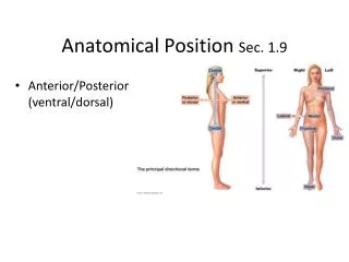
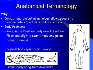
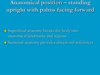
Anatomical position – standing upright with palms facing forward. Superficial anatomy breaks the body into anatomical landmarks and regions Sectional anatomy provides directional references. Figure 1.7 Anatomical Landmarks. Figure 1.7a. Figure 1.7b.

E N D
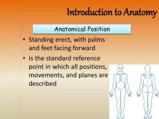
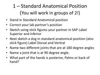
Anatomical position – standing upright with palms facing forward • Superficial anatomy breaks the body into anatomical landmarks and regions • Sectional anatomy provides directional references
Figure 1.7 Anatomical Landmarks Figure 1.7a
Figure 1.8 Abdominopelvic Quadrants and Regions Figure 1.8a
Figure 1.9 Directional References Figure 1.9
Planes and Sections are important in visualizing structures • Transverse plane divides the body into superior and inferior • Frontal (coronal) plane divides the body into anterior and posterior • Sagittal plane divides the body into left and right • Midsagittal divides the body exactly down the middle
Figure 1.10 Planes of Section Figure 1.10
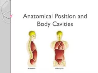
Body Cavities • Body cavities are internal chambers holding vital organs • Cavities protect vital organs • Cavities allow organs to change in shape and size • Two body cavities • Dorsal body cavity includes the cranial cavity and the spinal cavity • Ventral body cavity includes the thoracic cavity and the abdominopelvic cavity
Figure 1.12a Body Cavities Figure 1.12a, b
Thoracic Cavities • The thoracic cavity contains the heart and lungs. • It is subdivided into the left and right pleural cavities and the mediastinum • Each pleural cavity contains one lung lined by the visceral and parietal pleura • The mediastinum contains the pericardium, another serous membrane that surrounds the heart PLAY Animation: Heart Dissection
Abdominopelvic Cavity • The abdominopelvic cavity is lined by the peritoneum • The abdominal cavity extends from the diaphragm to the superior margins of the pelvis • liver, stomach, spleen and most of the large intestine
Abdominopelvic Cavity • The pelvic cavity is bordered by the pelvis, with a floor of muscle • reproductive organs, urinary bladder and the final portion of the large intestine PLAY Animation: Digestive System Dissection
Clinical technology allows many different views of the body • X-rays • Computerized tomography (CT) scans • Magnetic resonance imaging (MRI) scans • Ultrasound images • Spiral CT scans • Digital subtraction angiography images (DSA) • Positron emission tomography (PET) scans
Figure 1.13 X-rays Figure 1.13
Figure 1.14 Common scanning techniques Figure 1.14
Figure 1.15 Special Scanning Methods Figure 1.15c
You should now be familiar with: • The characteristics of life. • The sciences of anatomy and physiology and their various subdivisions. • The levels of organization in the human body. • The definition and importance of homeostasis. • The terminology associated with superficial and sectional anatomy and the body cavities.
Tissues and tissue types • Tissues are: • Collections of specialized cells and cell products organized to perform a limited number of functions • Histology = study of tissues • The four tissue types are: • Epithelial • Connective • Muscular • Nervous
Epithelial tissue • Includes glands and epithelium • Glands are secretory • Is avascular • Forms a protective barrier that regulates permeability • Cells may show polarity
Functions of epithelium • Physical protection • Control permeability • Provide sensation • Produce specialized secretions
Specializations of epithelium • Perform secretory functions • Perform transport functions • Maintain physical integrity • Ciliated epithelia move materials across their surface
Maintaining the integrity of epithelium • Cells attach via cell adhesion molecules (CAM) • Cells attach at specialized cell junctions • Tight junctions • Desmosomes • Gap junctions
Structure of typical epithelium • Basal lamina attaches to underlying surface • Lamina lucida • Lamina densa • Germinative cells replace short-lived epithelial cells
Classification of epithelia • Number of cell layers • Simple • Stratified • Shape of apical surface cells • Squamous • Cuboidal • Columnar
Glandular epithelia • Exocrine glands • Secrete through ducts onto the surface of the gland • Endocrine glands • Release hormones into surrounding fluid
Glandular secretions can be: • Merocrine (product released through exocytosis) • Apocrine (involves the loss of both product and cytoplasm) • Holocrine (destroys the cell)
Glands • Unicellular • Individual secretory cells • Multicellular • Organs containing glandular epithelium • Classified according to structure
Connective tissue functions: • Establishing a structural framework • Transporting fluids and dissolved materials • Protecting delicate organs • Supporting, surrounding and interconnecting tissues • Storing energy reserves • Defending the body from microorganisms
Connective tissues contain • Specialized cells • Matrix • Composed of extracellular protein fibers and a ground substance
Connective tissue proper • Contains varied cell populations • Contains various fiber types • A syrupy ground substance
Fluid connective tissue • Contains a distinctive cell population • Watery ground substance with dissolved proteins • Two types • Blood • Lymph
Supporting connective tissues • Less diverse cell population • Dense ground substance • Closely packed fibers • Two types • Cartilage • Bone
Connective tissue proper • Contains fibers, a viscous ground substance, and a varied cell population. • The ground substance is the non-living material in which the cells and protein fibres are found. • Can contain varying amounts of water. • Can be of viscous (blood), semi-solid (cartilage) or solid (bone). • The ground substance and the extracellular proteins form the matrix.
Types of cells found in connective tissue: • Macrophage • Adipocytes • Mesenchymal cells • Fibroblasts • Melanocytes • Mast cells • Lymphocytes • Microphages
Connective tissue proper • Three types of fiber • Collagen fibers • Reticular fibers • Elastic fibers
Connective tissue proper • Classified as loose or dense • Loose • Embryonic mesenchyme, mucous connective tissues • Areolar tissue • Adipose tissue • Reticular tissue • Dense • Dense regular CT • Dense irregular CT