Download
1 / 64
690 likes | 1.39k Vues
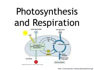
Respiration & Gas Exchange. Gas exchange is the uptake of molecular oxygen (O 2 ) from the environment and the discharge of carbon dioxide (CO 2 ) to the environment. While often called respiration, this process is distinct from, but linked to, the production of ATP in cellular respiration.

E N D
Gas exchange is the uptake of molecular oxygen (O2) from the environment and the discharge of carbon dioxide (CO2) to the environment. While often called respiration, this process is distinct from, but linked to, the production of ATP in cellular respiration. 1. Gas exchange supplies oxygen for cellular respiration and disposes of carbon dioxide: an overview
Gas exchange, in concert with the circulatory system, provide the oxygen necessary for aerobic cellular respiration and removes the waste product, carbon dioxide. Fig. 42.18
The source of oxygen, the respiratory medium, is air for terrestrial animals and water for aquatic animals. The atmosphere is about 21% O2 (by volume). Dissolved oxygen levels in lakes, oceans, and other bodies of water vary considerably, but they are always much less than an equivalent volume of air.
The part of an animal where gases are exchanged with the environment is the respiratory surface. Movements of CO2 and O2 across the respiratory surface occurs entirely by diffusion. The rate of diffusion is proportional to the surface area across which diffusion occurs, and inversely proportional to the square of the distance through which molecules must move. Therefore, respiratory surfaces tend to be thin and have large areas, maximizing the rate of gas exchange. In addition, the respiratory surface of terrestrial and aquatic animals are moist to maintain the cell membranes and thus gases must first dissolve in water.
Because the respiratory surface must supply O2 and expel CO2 for the entire body, the structure of a respiratory surface depends mainly on the size of the organism, whether it lives in water or on land, and by its metabolic demands. An endotherm has a larger area of respiratory surface than a similar-sized ectotherm.
Gas exchange occurs over the entire surface area of protists and other unicellular organisms. Similarly, for some relatively simple animals, such as sponges, cnidarians, and flatworms, the plasma membrane of every cell in the body is close enough to the outside environment for gases to diffuse in and out. However, in most animals, the bulk of the body lacks direct access to the respiratory medium. The respiratory surface is a thin, moist epithelium, separating the respiratory medium from the blood or capillaries, which transport gases to and from the rest of the body.
Some animals, such as earthworms and some amphibians, use the entire outer skin as a respiratory organ. Just below the moist skin is a dense net of capillaries. However, because the respiratory surface must be moist, their possible habitats are limited to water or damp places.
Animals that use their moist skin as their only respiratory organ are usually small and are either long and thin or flat in shape, with a high ratio of surface area to volume. For most other animals, the general body surface lacks sufficient area to exchange gases for the entire body. The solution is a respiratory organ that is extensively folded or branched, enlarging the surface area for gas exchange. Gills, tracheae, and lungs are the three most common respiratory organs.
Gills are outfoldings of the body surface that are suspended in water. The total surface area of gills is often much greater than that of the rest of the body. 2. Gills are respiratory adaptation of most aquatic animals
In some invertebrates, such as sea stars, the gills have a simple shape and are distributed over much of the body. Many segmented worms have flaplike gills that extend from each body segment, or long feathery gills clustered at the head or tail. The gills of clams, crayfish, and many other animals are restricted to a local body region. Fig. 42.19
Water has both advantages and disadvantages as a respiratory medium. There is no problem keeping the cell membranes of the respiratory surface moist, since the gills are surrounded by the aqueous environment. However, O2 concentrations in water are low, especially in warmer and saltier environments. Thus, gills must be very effective to obtain enough oxygen.
Ventilation, which increases the flow of the respiratory medium over the respiratory surface, ensures that there is a strong diffusion gradient between the gill surface and the environment. Without ventilation, a region of low O2 and high CO2 concentrations can form around the gill as it exchanges gas with the environment. Crayfish and lobsters have paddlelike appendages that drive a current of water over their gills.
Fish gills are ventilated by a current of water that enters the mouth, passes through slits in the pharynx, flows over the gills, and exits the body. Because water is dense and contains little oxygen per unit volume, fishes must expend considerable energy in ventilating their gills. Gas exchange at the gill surface is enhanced by the opposing flows of water and blood at the gills.
This flow pattern is countercurrent exchange. As blood moves anteriorly in a gill capillary, it becomes more and more loaded with oxygen, but it simultaneously encounters water with even higher oxygen concentrations because it is just beginning its passage over the gills. All along the gill capillary, there is a diffusion gradient favoring the transfer of oxygen from water to blood. Fig. 42.20
Gills are generally unsuited for an animal living on land. An expansive surface of wet membrane exposed to air would lose too much water by evaporation. In addition, the gills would collapse as their fine filaments, no longer supported by water, would cling together, reducing surface area for exchange. Most terrestrial animals have their respiratory surfaces within the body, opening to the atmosphere through narrow tubes.
As a respiratory medium, air has many advantages over water. Air has a much higher concentration of oxygen. Also, since O2 and CO2 diffuse much faster in air than in water, respiratory surfaces exposed to air do not have to be ventilated as thoroughly as gills. When a terrestrial animal does ventilate, less energy is needed because air is far lighter and much easier to pump than water and much less volume needs to be breathed to obtain an equal amount of O2. 3. Tracheal systems and lungs are respiratory adaptations of terrestrial animals
Air does have problems as a respiratory medium. The respiratory surface, which must be large and moist, continuously loses water to the air by evaporation. This problem is greater reduced by a respiratory surface folded into the body.
The tracheal system of insects is composed of air tubes that branch throughout the body. The largest tubes, called tracheae, open to the outside, and the finest branches extend to the surface of nearly every cell where gas is exchanged by diffusion across the moist epithelium that lines the terminal ends. The open circulatory system does not transport oxygen and carbon dioxide.
For a small insect, diffusion through the trachea brings in enough O2 and removes enough CO2 to support cellular respiration. Larger insects with higher energy demands ventilate their tracheal systems with rhythmic body movements that compress and expand the air tubes like bellows. An insect in flight has a very high metabolic rate, consuming 10 to 200 times more O2 than it does at rest. Alternating contraction and relaxation of flight muscles compress and expand the body, rapidly pumping air through the tracheal system. The flight muscles are packed with mitochondria, and the tracheal tubes supply each with amply oxygen.
Unlike branching tracheal systems, lungs are restricted to one location. Because the respiratory surface of the lung is not in direct contact with all other parts of the body, the circulatory system transports gases between the lungs and the rest of the body. Lungs have a dense net of capillaries just under the epithelium that forms the respiratory surface. Lungs have evolved in spiders, terrestrial snails, and vertebrates.
Among the vertebrates, amphibians have relatively small lungs that do not provide a large surface (many lack lungs altogether). They rely heavily on diffusion across other body surfaces, especially their moist skin, for gas exchange. In contrast, most reptiles and all birds and mammals rely entirely on lungs for gas exchange. Turtles may supplement lung breathing with gas exchange across moist epithelial surfaces in their mouth and anus. Lungs and air-breathing have evolved in a few fish species as adaptations to living on oxygen-poor water or to spending time exposed to air.
Located in the thoracic (chest) cavity, the lungs of mammals have a spongy texture and are honeycombed with a moist epithelium that functions as the respiratory surface. A system of branching ducts conveys air to the lungs.
Air enters through the nostrils and is then filtered by hairs, warmed and humidified, and sampled for odors as it flows through the nasal cavity. The nasal cavity leads to the pharynx, and when the glottis is open, air enters the larynx, the upper part of the respiratory tract. The wall of the larynx is reinforced by cartilage. In most mammals, the larynx is adapted as a voicebox in which vibrations of a pair of vocal cords produce sounds These sounds are high-pitched when the the cords are stretched tight and vibrate rapidly and low-pitched when the cords are less tense and vibrate slowly.
From the larynx, air passes into the trachea, or windpipe, whose shape is maintained by rings of cartilage. The trachea forks into two bronchi, one leading into each lung. Within the lung, each bronchus branches repeatedly into finer and finer tubes, called bronchioles. The epithelium lining the major branches of the respiratory tree is covered by cilia and a thin film of mucus. The mucus traps dust, pollen, and other particulate contaminants, and the beating cilia move the mucus upward to the pharynx, where it is swallowed.
At their tips, the tiniest bronchioles dead-end as a cluster of air sacs called alveoli. Gas exchange occurs across the thin epithelium of the lung’s millions of alveoli. These have a total surface area of about 100 m2 in humans. Oxygen in the air entering the alveoli dissolves in the moist film and rapidly diffuses across the epithelium into a web of capillaries that surrounds each alveolus. Carbon dioxide diffuses in the opposite direction.
The process of breathing, the alternate inhalation and exhalation of air, ventilates lungs. A frog ventilates its lungs by positive pressure breathing. During a breathing cycle, muscles lower the floor of the oral cavity, enlarging it and drawing in air through the nostrils. With the nostrils and mouth closed, the floor of the oral cavity rises and air is forced down the trachea. Elastic recoil of the lungs, together with compression of the muscular body wall, forces air back out of the lungs during exhalation.
In contrast, mammals ventilate their lungs by negative pressure breathing. This works like a suction pump, pulling air instead of pushing it into the lungs. Muscle action changes the volume of the rib cage and the chest cavity,and the lungsfollow suit. Fig. 42.24
The lungs are enclosed by a double-walled sac, with the inner layer of the sac adhering to the outside of the lungs and the outer layer adhering to the wall of the chest cavity. A thin space filled with fluid separates the two layers. Because of surface tension, the two layers behave like two plates of glass stuck together by the adhesion and cohesion of a film of water. The layers can slide smoothly past each other, but they cannot be pulled apart easily. Surface tension couples movements of the lungs to movements of the rib cage.
Lung volume increases as a result of contraction of the rib muscles and diaphragm, a sheet of skeletal muscle that forms the bottom wall of the chest cavity. Contraction of the rib muscles expands the rib cage by pulling the ribs upward and the breastbone outward. At the same time, the diaphragm contracts and descends like a piston. These changes increase the lung volume, and as a result, air pressure within the alveoli becomes lower than atmospheric pressure. Because air flows from higher pressure to lower pressure, air rushes into the respiratory system.
During exhalation, the rib muscles and diaphragm relax. This reduces lung volume and increases air pressure within the alveoli. This forces air up the breathing tubes and out through the nostrils.
Actions of the rib muscles and diaphragm accounts for changes in lung volume during shallow breathing, when a mammal is at rest. During vigorous exercise, other muscles of the neck, back, and chest further increase ventilation volume by raising the rib cage even more. In some species, rhythmic movements during running cause visceral organs, including the stomach and liver, to slide forward and backward in the body cavity with each stride. This “visceral pump” further increases ventilation volume by adding to the piston-like action of the diaphragm.
The volume of air an animal inhales and exhales with each breath is called tidal volume. It averages about 500 mL in resting humans. The maximum tidal volume during forced breathing is the vital capacity, which is about 3.4 L and 4.8 L for college-age females and males, respectively. The lungs hold more air than the vital capacity, but some air remains in the lungs, the residual volume, because the alveoli do not completely collapse.
Since the lungs do not completely empty and refill with each breath cycle, newly inhaled air is mixed with oxygen-depleted residual air. Therefore, the maximum oxygen concentration in the alveoli is considerably less than in the atmosphere. This limits the effectiveness of gas exchange.
Ventilation is much more complex in birds than in mammals. Besides lungs, birds have eight or nine air sacs that do not function directly in gas exchange, but act as bellows that keep air flowing through the lungs. Fig. 42.25
The entire system - lungs and air sacs - is ventilated when the bird breathes. Air flows through the interconnected system in a circuit that passes through the lungs in one direction only, regardless of whether the bird is inhaling or exhaling. Instead of alveoli, which are dead ends, the sites of gas exchange in bird lungs are tiny channels called parabronchi, through which air flows in one direction.
This system completely exchanges the air in the lungs with every breath. Therefore, the maximum lung oxygen concentrations are higher in birds than in mammals. Partly because of this efficiency advantage, birds perform much better than mammals at high altitude. For example, while human mountaineers experience tremendous difficulty obtaining oxygen when climbing the Earth’s highest peaks, several species of birds easily fly over the same mountains during migration.
While we can voluntarily hold our breath or breath faster and deeper, most of the time autonomic mechanisms regulate our breathing. This ensures that the work of the respiratory system is coordinated with that of the cardiovascular system, and with the body’s metabolic demands for gas exchange. 4. Control centers in the brain regulate the rate and depth of breathing
Our breathing control centers are located in two brain regions, the medulla oblongata and the pons. Aided by the control center in the pons, the medulla’s center sets basic breathing rhythm, triggering contraction of the diaphragm and rib muscles. A negative-feedback mechanism via stretch receptors prevents our lungs from overexpanding by inhibiting the breathing center in the medulla.
The medulla’s control center monitors the CO2 level of the blood and regulated breathing activity appropriately. Its main cues about CO2 concentration come from slight changes in the pH of the blood and cerebrospinal fluid bathing the brain. Carbon dioxide reacts with water to form carbonic acid, which lowers the pH. When the control center registers a slight drop in pH, it increases the depth and rate of breathing, and the excess CO2 is eliminated in exhaled air.
Oxygen concentrations in the blood usually have little effect of the breathing control centers. However, when the O2 level is severely depressed - at high altitudes, for example, O2 sensors in the aorta and carotid arteries in the neck send alarm signals to the breathing control centers, which respond by increasing breathing rate. Normally, a rise in CO2 concentration is a good indicator of a fall in O2 concentrations, because these are linked by the same process - cellular respiration. However, deep, rapid breathing purges the blood of so much CO2 that the breathing center temporarily ceases to send impulses to the rib muscles and diaphragm.
The breathing center responds to a variety of nervous and chemical signals and adjusts the rate and depth of breathing to meet the changing demands of the body. However, breathing control is only effective if it is coordinated with control of the circulatory system, so that there is a good match between lung ventilation and the amount of blood flowing through alveolar capillaries. For example, during exercise, cardiac output is matched to the increased breathing rate, which enhances O2 uptake and CO2 removal as blood flows through the lungs.
For a gas, whether present in air or dissolved in water, diffusion depends on differences in a quantity called partial pressure, the contribution of a particular gas to the overall total. At sea level, the atmosphere exerts a total pressure of 760 mm Hg. Since the atmosphere is 21% oxygen (by volume), the partial pressure of oxygen (abbreviated PO2) is 0.21 x 760, or about 160 mm Hg. The partial pressure of CO2 is only 0.23 mm Hg. 5. Gases diffuse down pressure gradients in the lungs and other organs
When water is exposed to air, the amount of a gas that dissolves in water is proportional to its partial pressure in the air and its solubility in water. An equilibrium is eventually reached when gas molecules enter and leave the solution at the same rate. At this point, the gas is said to have the same partial pressure in the solution as it does in the air. Thus, in a glass of water exposed to air at sea-level air pressure, the PO2 is 160 mm Hg and the PCO2 is 0.23 mm Hg. A gas will always diffuse from a region of higher partial pressure to a region of lower partial pressure.
Blood arriving at the lungs via the pulmonary arteries has a lower PO2 and a higher PCO2 than the air in the alveoli. As blood enters the alveolar capillaries, CO2 diffuses from blood to the air within the alveoli, and oxygen in the alveolar air dissolves in the fluid that coats the epithelium and diffuses across the surface into the blood. By the time blood leaves the lungs in the pulmonary veins, its PO2 have been raised and its PCO2 has been lowered.
In the tissue capillaries, gradients of partial pressure favor the diffusion of oxygen out of the blood and carbon dioxide into the blood. Cellular respiration removes oxygen from and adds carbon dioxide to the interstitial fluid by diffusion, and from the mitochondria in nearby cells. After the blood unloads oxygen and loads carbon dioxide, it is returned to the heart and pumped to the lungs again, where it exchanges gases with air in the alveoli.