Download
1 / 27
290 likes | 726 Vues
PURIFICATION AND CHARACTERIZATION OF PROTEINS. BY ROHAN MENON. INTRODUCTION. Goals of purification vary with the intended use of the protein.

E N D
PURIFICATION AND CHARACTERIZATION OF PROTEINS BY ROHAN MENON
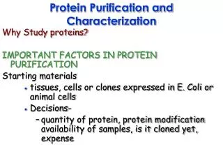
INTRODUCTION • Goals of purification vary with the intended use of the protein. • Purity is defined by the general level of protein contaminants and also by the absence of contaminants of special interest such as endotoxin, viruses etc. • Protein purification can be divided into 5 stages. a) Preparation of the source b) Knowledge of protein properties c) Development of an Assay d) Primary Isolation e) Final Purification
PREPARATION OF SOURCE • Selection of raw materials from which proteins will be isolated (microbial or cultured metazoan cell line). • Protein supplies can be increased by increasing the cultivation volume ( by growing more cells per unit volume).
KNOWLEDGE OF PROTEIN PROPERTIES • Source: cell type, intra/ extra cellular location, folding state, presence of proteases / glycosidases • Stability: to temperature range, pH range, ionic strength, hydrophobic surfaces, aggregation tendency, cofactor of metal ion loss / requirement. • Size : molecular weight, peptide chain (s), hydrodynamic radius. • Charge: isoelectric point, titration curve, electrophoretic mobility. • Binding partners: substrates and cofactors, screening-derived binding agents, metal affinity.
ASSAY • An assay for the desired activity or protein is required. • They must be convenient , rapid and extremely precise.
INITIAL ISOLATION This mainly consists of separation of proteins from water and other cell components. a) Concentration b) Cell lysis c) Refolding
CONCENTRATION • Extra cellular proteins are usually concentrated from the cell by ultra filtration or adsorption. • Secreted protein adsorbed to the outside of the cell and can be concentrated along with them and then liberated by washing, often with a high salt buffer.
CELL LYSIS • Intra cellular proteins are liberated by cell lysis. • Cell lysis is the process of disintegration of a cell (French Press – forcing cell through an orifice at high pressure). • Soluble proteins are often recovered from cell lysates by precipitation with ammonium sulfate or polyethylene glycol.
REFOLDING • Recombinant proteins often misfold to form dense, insoluble aggregates of inactive protein. • The first step in the renaturation process is the dissolution of the inclusion bodies in a strong chaotrope solution with 6M urea. Dissolution in denaturant is rapid and reliable. • The denatured protein is then allowed to renature to its native confirmation by removing the denaturant through dialysis, dilution or chromatographic separation. Allow the refolding process for 7 to 10 days.
HIGH RESOLUTION PURIFICATION • Chromatography is the usual method of preparing highly purified active proteins. • Chromatographic operations are classified as low-pressure, medium-pressure, high-pressure depending on the pressure used to force liquid through the packed bed.
HIGH RESOLUTION PURIFICATION • Chromatographic operations are broadly classified as a) Ion – exchange Chromatography b) Hydrophobic Chromatography c) Affinity Chromatography d) Size exclusion Chromatography
Ion – exchange Chromatography In this case, a cation (or alternatively an anion) is attached to the resin beads. Depending upon the electrical properties of the proteins, they may attach to the column. For example, positively charged proteins will stick to a negatively charged column. These proteins can then be removed by washing the column with either a strong salt solution or changing the pH of the wash buffer. • Anion exchangers such as DEAE ( Diethyl amino ethyl) are used. Attraction of proteins at a pH above the isolectric point of the protein. • Cation exchangers such as CM ( Carboxy methyl) are used. Attraction of protein at a pH below the isoelectric point of the protein.
ELUTION • Done by washing the column with a strong salt solution (NaCl) which increases the ionic strength thereby pushing out the proteins.
HYDROPHOBIC CHROMATOGRAPHY • Principle Proteins are separated by hydrophobic interaction on columns with hydrophobic groups attached (e.g. phenyl-, octyl groups) • Surface hydrophobicity Hydrophobicity of amino acid sidechains Tryptofan > Isoleucine, Phenylalanine > Tyrosine > Leucine > Valine > Methionine Most hydrophobic sidechains are buried in interior of protein, but some (clusters of) hydrophobic groups occur at surface of protein. Surface hydrophobic sidechains can interact with hydrophobic groups for example attached to a column.
HYDROPHOBIC CHROMATOGRAPHY • Temperature Increasing temperature --> stronger hydrophobic interactions • Sample (application) Column having high concentration of a salt promotes binding (for example ammonium sulfate just below the concentration that starts to precipitate protein). • Elution of bound proteins Negative gradient of salting-out ions (from high to low concentration).
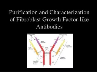
AFFINITY CHROMATOGRAPHY • In this type of chromatography, a compound with a special affinity for the protein of interest is attached to the resin. For example, in immunoaffinity chromatography antibodies to a specific protein (or its domain) are used as the specialised compound. • The resin is then packed into a column. When a mixture of proteins is passed through the column, only those proteins with special affinity for the compound will stick to the column. All the other proteins will pass through the column. Once the non-specific proteins are eluted, proteins of interest that have stuck to the column can be eluted. These proteins can be removed by changing the ionic strength of the solution (so affecting the strength of binding of the protein to the column). Alternatively the special compound can be added to the elution solution and the equilibrium will change so that the protein will no longer stick to the column.
SIZE EXCLUSION CHROMATOGRAPHY • It is also known as Gel Filtration • Used to separate proteins on the basis of their molecular weight. The column is packed with a porous resin. • The matrix retards proteins of different sizes for different periods. The proteins are collected automatically as they flow out of the column in tubes held in a fraction collector. • Larger proteins will be eluted first since the smaller proteins travel through the pores of the resin.
CHARACTERIZATION The method of protein characterization are as follows • Electrophoresis • Peptide Sequencing • Tryptic Mapping • Analytical Ultracentrifugation • Spectroscopy • Biosensors • Mass Spectrometry
ELECTROPHORESIS • Proteins are separated on the basis of their molecular mass using sodium dodecyl sulfate polyacrylamine (SDS-PAGE). • It reduces proteins into regular rod like forms of constant charge density per unit mass. • SDS breaks all hydrogen bonds and partially unfolds the protein structure. • The other method coming up is Capillary electrophoresis. • Capillary electrophoresis a) Conducted in a tubing of very small diameter using high voltage. b) Takes very less time. c) Good separation is achieved.
PEPTIDE SEQUENCING • Also known as amino terminal sequencing. • Used to identify the first few amino acids of the protein. • This sequence information can be used to confirm the identity of the protein. • It depends on sequential stepwise removal of N-terminal amino acids by HPLC and then identified by characteristic retention times. TRYPTIC MAPPING • Small peptides derived from the protein by endoprotease action are separated by high resolution reverse phase HPLC. • The individual peptides are then subjected to sequencing to confirm the identity of the given peptide.
ANALYTICAL ULTRACENTRIFUGATION • This technique allows measurement of a variety of properties of a protein sample, including solution molecular weight, interaction with other molecules and sample homogeneity. SPECTROSCOPY • It gives an indication of the fraction of the polypeptide that is composed of specific secondary structural features such as α-helix and β-sheet. • It is also used for characterizing metal containing protein cofactors.
MASS SPECTROMETRY BIOSENSORS • Device used for the detection of protein cells. • An antibody to a particular protein is immobilized on the sensor surface, addition of sample containing that particular protein will produce an immediate signal. • A technique by which you can determine the mass of a protein with remarkable precision of the order of a few hydrogen atoms. • It can detect any important modification (post translational modification) or variation in a protein structure.
EMERGING TRENDS • Automation • Capillary Electrophoresis and Mass spectrometry are advancing rapidly. Mass Spectrometry will be more widely used, largely through core facilities. CE will likely replace SDS-PAGE in a few more years.