Download
1 / 22
270 likes | 396 Vues
Explore protein purification techniques like salting out, differential centrifugation, and column chromatography to isolate proteins from cells. Learn electrophoresis and sequencing methods for protein characterization.

E N D
Chapter FiveProtein Purification and Characterization Techniques
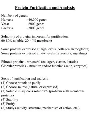
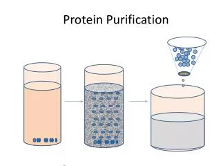
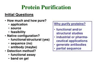
Isolation of Proteins from Cells Many different proteins exists within one cell • Many steps needed to extract protein of interest, and separate from many contaminants • Before purification begins, protein must be released from cell by homogenization
Salting Out • After Proteins solubilized, they can be purified based on solubility (usually dependent on overall charge, ionic strength, polarity • Ammonium sulfate (NH4SO4) commonly used to “salt out” • Takes away water by interacting with it, makes protein less soluble because hydrophobic interactions among proteins increases • Different aliquots taken as function of salt concentration to get closer to desired protein sample of interest (30, 40, 50, 75% increments) • One fraction has protein of interest
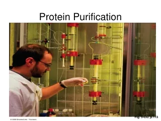
Differential Centrifugation • Sample is spun, after lysis, to separate unbroken cells, nuclei, other organelles and particles not soluble in buffer used • Different speeds of spin allow for particle separation
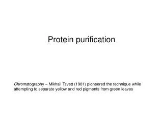
Column Chromatography • Basis of Chromatography • Different compounds distribute themselves to a varying extent between different phases • Interact/distribute themselves • In different phases • 2 phases: • Stationary: samples interacts with this phase • Mobile: Flows over the stationary phase and carries along with it the sample to be separated
Size-Exclusion/Gel-Filtration • Separates molecules based on size. • Stationary phase composed of cross-linked gel particles. • Extent of cross-linking can be controlled to determine pore size • Smaller molecules enter the pores and are delayed in elution time. Larger molecules do not enter and elute from column before smaller ones.
Affinity Chromatography • Uses specific binding properties of molecules/proteins • Stationary phase has a polymer that can be covalently linked to a compound called a ligand that specifically binds to protein
Ion Exchange • Interaction based on overall charge (less specific than affinity) • Cation exchange • Anion exchange
Electrophoresis • Electrophoresis- charged particles migrate in electric field toward opposite charge • Proteins have different mobility: • Charge • Size • Shape • Agarose used as matrix for nucleic acids • Polyacrylamide used mostly for proteins
Electrophoresis (Cont’d) • Polyacrylamide has more resistance towards larger molecules than smaller • Protein is treated with detergent (SDS) sodium dodecyl sulfate • Smaller proteins move through faster (charge and shape usually similar)
Isoelectric Focusing • Isolectric focusing- based on differing isoelectric pts. (pI) of proteins • Gel is prepared with pH gradient that parallels electric-field. What does this do? • Charge on the protein changes as it migrates. • When it gets to pI, has no charge and stops
Primary Structure Determination How is 1˚ structure determined? • Determine which amino acids are present (amino acid analysis) • Determine the N- and C- termini of the sequence (a.a sequencing) • Determine the sequence of smaller peptide fragments (most proteins > 100 a.a) 4) Some type of cleavage into smaller units necessary
Protein Cleavage Protein cleaved at specific sites by: • Enzymes- Trypsin, Chymotrypsin • Chemical reagents- Cyanogen bromide Enzymes: Trypsin- Cleaves @ C-terminal of (+) charged side chains Chymotrypsin- Cleaves @ C-terminal of aromatics
Cleavage by CnBr Cleaves @ C-terminal of INTERNAL methionines
Determining Protein Sequence After cleavage, mixture of peptide fragments produced. • Can be separated by HPLC or other chromatographic techniques • Use different cleavage reagents to help in 1˚ determination
Peptide Sequencing • Can be accomplished by Edman Degradation • Relatively short sequences (30-40 amino acids) can be determined quickly • So efficient, today N-/C-terminal residues usually not done by enzymatic/chemical cleavage