Download
1 / 39
420 likes | 1.11k Vues
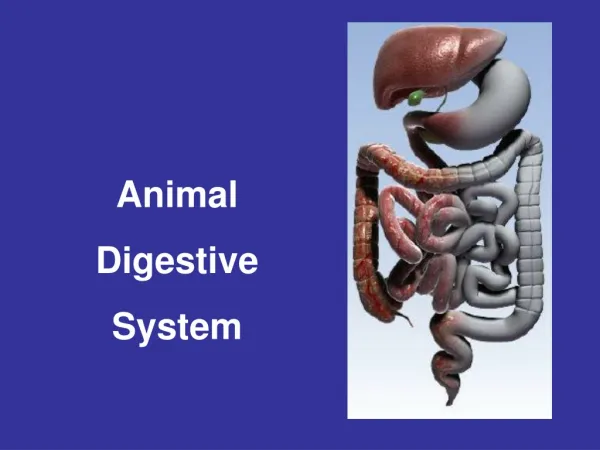
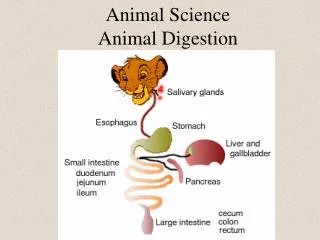
Animal Science Animal Digestion. Objective 1 Name, locate and describe the functions of the parts of the digestive systems of ruminant and nonruminant animals. Beef: Mouth- point at which ingestion takes place and where cud is chewed. Also, the body is stimulated to secrete saliva.

E N D
Objective 1Name, locate and describe the functions of the parts of the digestive systems of ruminant and nonruminant animals. • Beef: • Mouth- point at which ingestion takes place and where cud is chewed. • Also, the body is stimulated to secrete saliva.
Teeth- provide mechanical digestion of feed by breaking, cutting, and tearing up food. • The increase surface area aids in the chewing and swallowing process.
Salivary Glands- • secrete saliva which moistens the feed and stimulates taste. • Saliva contains the enzymes, • Enzymes are organic catalysts that speed up chemical reactions without being altered by the reaction.
Saliva includes: • Water: moistens consumed feed and aids in the taste mechanisms. • Mucin: lubrication aid for swallowing. • Bicarbonate Salts: acts as a buffer to regulate pH of the stomach. • Enzyme: salivary amylase initiates carbohydrate breakdown. • Mature horses can produce 10 gal/day; cows- 12 gal/day; and sheep- 2 gal/day.
Esophagus: hollow muscular tube that transports ingesta from the mouth to the stomach; • Ingesta material is moved by a series of muscular contractions referred to as peristatic waves. • Also serves as a storage for food (crop) in chickens. • Cardiac Sphincter: valve at the junction of the stomach and esophagus.
Stomach: hollow, pear shaped , muscular digestive organ. Functions: (Sight of Digestion) - Storage of ingested feed. - Muscular movements causing physical breakdown. - Secretes digestive juices: 1) Hydrochloric Acid 2) Pepsin 3) Rennin
Stomach cont. • Stomach contents approximately a pH of 2 (bacterial effect). • Material leaving the stomach is called chyme.
Parts of the stomach-simple stomach • Cardia sphincter- see above • Esophageal region: non-glandular area surrounding the cardia • Cardiac gland region: contains cells that produce primarily mucus (protects stomach lining)
Fundic gland region: contains cells that provide the gastric secretions needed for the initial stages of digestion. • Parietal cells: produce hydrochloric acid. • Chief cells: produce enzymes or precursors of enzymes • Pyloric gland region: contains cells that produce mucus and some proteolytic enzymes. • Pylorus sphincter: at the beginning of the small intestine which controls passage of material (chyme) out of the stomach
Small Intestine • Divided into 3 sections. • Duodenum: first sections • Receives secretions from: • Pancreas: acts on proteins, carbohydrates and lipids • Liver: bile (stored in the gallbladder) breaks down fat. *Horses do not have gallbladders • Active site of digestion • Jejunum: middle section; active in nutrient absorption • Ileum: last section; active in nutrient absorption
Small Intestines • Walls of the S.I. are lined with a series of fingerlike projections called villi, which in turn have minute projections called microvillithat increase the nutrient absorption area.
Each nutrient has a specific absorption site. Each villus contains an arteriole and venule, together with a drainage tube of the lymphatic system, a lacteal. • The venules ultimately drain into the portal blood system, which goes directly to the liver; the lymph system empties via the thoracic duct into the vena cava.
S.I. contents are approximately pH 6 to 7. • Sight of digestion and absorption. • Passive absorption = results from diffusion or movement from high concentrations to low concentrations. • Active absorption = transport of molecules across the intestinal lining. • (villi) engulf the molecules and then actively transport these molecules to either the bloodstream or the lymph. • Feed is carried to the liver where it is detoxified.
Large Intestine • Divided into 3 sections • Cecum: • first sections; • size varies considerably in different species; • little functional significance in the pig. • Horses contain an active flora of bacteria similar to the microbial population in rumen compartment of ruminants. • Bacterial breakdown of cellulose and other carbohydrate material to produce VFAs (acetic, propionic and butyric) thus, the horse can utilize fibrous feeds. • Site of bacterial synthesis of water-soluble vitamins and protein.
Colon: • middle section; • largest part of the L.I. • Primary area of water restoration from intestinal contents. • Rectum: last section of the L.I. and the end of the digestive tract before the unabsorbed material (feces) is excreted out the anus.
Functions of the L.I. • Site of water restoration • Secretion of some mineral elements • calcium • Storage reservoir of undigested GIT contents. • Bacterial fermentation: • Synthesis of some water-soluble vitamins and vitamin K. • Some bacterial breakdown of fibrous ingredients. • Synthesis of some protein • Limited absorption of feedstuff from the L.I.
Anus: eternal opening where unabsorbed materials (feces) are expelled from the body.
Beef Cattle • Teeth: • Designed to shred fibrous material on the side of the mouth. • Saliva: • Contains no enzymes • Provides source of N (urea), P, Na • Utilized by rumen microorganisms • Aids in maintaining an appropriate pH in the rumen.
Beef Cattle Stomach • Divided into 4 compartments • Reticulum • Rumen • Omasum • Abomasum
Reticulum (Honeycomb) • First compartment and not completely separated from the rumen; • esophagus opening (cardia) is common to both reticulum and rumen compartments. • Walls are lined with mucus membrane containing many intersecting ridges that subdivide the surface into honeycomb-like compartment; • this wall arrangement traps hardware (nails, wire, and etc.) and does not allow it to proceed through the remainder of the GIT (gastrointestinal tract)
Reticulum (Honeycomb) • The wall does not secrete any enzymes. • The reticulum functions in moving ingested feed into the rumen or into the omasum and in regurgitation of ingesta during rumination.
Rumen (paunch) • Large, hollow, muscular compartment that extends from the diaphragm to the pelvis and almost entirely fills the left side of the abdominal cavity. • The wall of the mature rumen contains small tongue like projections called papillae, which can be readily identified by the naked eye. The walls aid in the secretion of enzymes.
Rumen (paunch) • Functions include: • Storage • Soaking • Physical mixing and breakdown.
Rumen (paunch) • Fermentation chamber: • ideal environment for microbial organisms • (bacterial and protozoa) • it is moist, warm, anaerobic, desirable in PH and there is an irregular introduction of new ingesta and a more or less continual removal of fermented digesta and end products of digestion. • Various types of bacteria are found with typical counts approaching numbers of 25 to 50 billion per milliliter of ruminal fluid.
Rumen (paunch) • This extensive pregastric fermentation result in: • Bacterial synthesis of water-soluble vitamins and vitamin K. • Bacterial synthesis of amino acids and protein. • As the bacteria move out of the rumen, they will be degraded in the lower portion of the host animal’s GIT and serve as a source of amino to the host animal.
Rumen (paunch) • Breakdown of fibrous feeds (high in cellulose). Bacteria contain enzymes to rupture cellulose bonding as well as starch bonding. • Rumen compartment is quite undeveloped at birth and may be functional by 6 to 8 weeks of age.
Omasum (many plies) • The omasum is a spherical organ filled with muscular laminae studded with short, blunt papillae. The walls secrete no enzymes. • The omasum is located to the right of the rumen and reticulum. • The omasum appears to be instrumental in reducing particle size of ingesta before it enters the abomasum and some absorption of water.
Abomasum (true or glandular stomach): • First glandular portion of the ruminant GIT (walls secrete enzymes). • Located below the omasum and extends caudally on the right side of the rumen. • In general, the gland regions of the abomasum correspond to the gland regions in the simple stomach of the nonruminant.
Other Peculiarities of the Ruminant Digestive System • Esophageal (or reticular) groove • Rumination • Eructation (belching of gas)
Esophageal (or reticular) groove • A passageway that extends from the cardia to the omasum, formed by two heavy muscular folds or lips, which can close to directly, or open and permit the ingesta to enter the rumen and reticulum. • Functions to allow milk consumed by the suckling animal to bypass the reticulorumen and escape bacterial fermentation. • Does not appear to remain in older animals.
Rumination • A process that permits an animal to forage and ingest feed rapidly, then complete the chewing at a later time; steps include regurgitation of feed, remastication, re-salivation and finally re-swallowing. • The regurgitation step is preceded by contraction of the reticulum; probably a reverse peristalsis in the esophagus is the major factor in moving the material up to the mouth, where excess liquid is squeezed out and swallowed.
Rumination cont. • The regurgitated material consists largely of roughage and fluid with little if any concentrate. • Cattle average about 8 hours per day ruminating. One rumination cycle requires about 1 minute, of which 3 to 4 seconds is utilized for both regurgitation and re-swallowing.
Eructation (belching of gas) • Microbial fermentation in the rumen results in production of large amounts of gases (primarily carbon dioxide and methane), which must be eliminated. • Contractions of the upper sacs of the rumen force gases forward and down; the esophagus then dilated and allows the gases to escape.
Eructation cont. • Bloat is a common problem in ruminants in which gas cannot escape. This creates a distention of the rumen, which can be seen on the left side of the animal; in most cases of bloat, a stable froth or foam is produced in the rumen. This interferes with normal belching and gas accumulates. Therefore, control measures must prevent foam or break it rapidly after it has formed. *ALL OTHER SYSTEMS ARE THE SAME.