Bioassay
it contains principle and application of bioassay, types of bioassay, bioassay of insulin,bioassay of oxytocin,bioassay of vasopressin, bioassay of ACTH,bioassay of of D- tubocurarine, bioassay of Digitalis, bioassay of Histamine and bioassay of 5-HT (serotonin).

Bioassay
E N D
Presentation Transcript
BIOassay Prepared By Ms. Prexita Patel Dept. of Pharmacology Anand Pharmacy College, Anand
Principle and Applications of Bioassay Definition:- Estimation of the potency of an active principle in a unit quantity of preparation or detection and measurement of the concentration of the substance in a preparation using biological methods (i.e. observation of pharmacological effects on living tissues, microorganisms or immune cells or animal) is known as Biological assay or Bioassay. Microbioassay, radioimmunoassay are also regarded as `bioassay'. Other synonyms are biometrics or biological standardization. Biological standardization cannot be considered as synonym of bioassay because in bioassay one simply estimates the potency in a preparation. However, in biological standardization, one has to increase or decrease the potency (concentration) of the sample/batch to bring to the standard level. Recently `biotechnology' has also been considered for bioassay. Bioassay of the products like erythropoietin, hepatitis- B vaccine etc. is being done through biotechnology.
Importance of Bioassay :- • Bioassays, as compared to other methods of assays (e.g. chemical or physical assay) are less accurate, less elaborate, more laborious, more troublesome and more expensive. Bioassay when carried out in whole animal, the effect of drug may be influenced by various factors like metabolism, nervous control; blood supply etc. cruelty of Animal Act is problem in bioassay. • Bioassay consider under following conditions:- • 1. Active principle of drug is unknown or cannot be isolated, e.g. insulin, streptokinase, urokinase etc. • 2. Chemical method is either not available or if available, it is too complex and insensitive or requires higher dose e.g. acetylcholine, glucagen. • 3. Chemical composition is not known, e.g. vaccines, antitoxins, toxins, and herbal preparation. • 4. Chemical composition of drug differs but have the same pharmacological action and vice-versa, e.g. cardiac glycosides, catecholamines etc. • The purpose of bioassay is to ascertain the potency of a drug and hence it serves as the quantitative part of any screening procedure (Research). Other purpose of bioassay is to standardize the preparation so that each contains the uniform specified pharmacological activity. In this way, it serves as a pointer in the Commercial Production of drugs when chemical assays are not available or do not suffice.
Principle of Bioassay:- The basic principle of bioassay is to compare the test substance with the International Standard preparation of the same and to find out how much test substance is required to produce the same biological effect, as produced by the standard. The standards are internationally accepted samples of drugs maintained and recommended by the Expert Committee of the Biological Standardization of W.H.O. They represent the fixed units of activity (definite weight of preparation) for drugs. In India, standard drugs are maintained in Government institutions like Central Drug Research Institute, Lucknow, Central Drug Laboratory, Calcutta, etc. The problem of biological variation must be minimized as far as possible. For that, one should keep uniform experimental conditions and assure the reproducibility of the responses. Statistical analysis P value) is commonly employed to biological variations.
Bioassay in Pharmacopoeia:- Pharmacopoeia gives guidelines for various assays including bioassays. The purpose of mentioning the bioassay method is to ensure the purity of material and determine total activity of a drug in container. For last two to three decades there has been a change in recognizing bioassay in pharmacopoeia. This change has been in parallel with rapid developments and advancements in analytical techniques. 2. Types of Bioassay:- There are two types of Bioassay: (I) – Methods of bioassay for Agonist. (II) – Methods of bioassay for Antagonist.
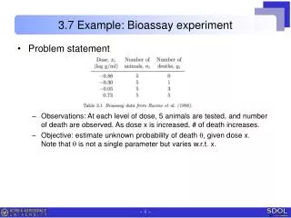
I. Methods of Bioassay for Agonist:- • An agonist may produce graded response or quantal response. • Graded response means that the response is proportional to the dose and response may lie between no response and the maximum response. • By quantal, it is meant that the response is in the form of "all or none", i.e. either no response or maximum response. • The drugs producing quantal effect can be bioassayed by end point method and the drugs producing graded responses can be bioassayed by • (1) Matching or bracketing method or (2) Graphical method. • End Point Method:- • Here the threshold dose producing a positive effect is measured on each animal and the comparison between the average results of two groups of animals (one receiving standard and other the test) is done. e.g. bioassay of digitalis in cats. • Here the cat is anaesthetized with chloralose and its blood pressure is recorded. The drug is then slowly infused into the animal and the moment the heart stops beating and blood pressure falls to zero, the volume of fluid infused is noted down.
Two series of such experiments-one using standard digitalis and the other using test preparation of digitalis is done and then potency is calculated as follows:
3. Rat diaphragm method:- In this method increase in glycogen content of the muscle or increase in glucose uptake by muscle in response to insulin is taken as the index of potency of insulin. 4. Rat epididymal fat-pad method: - Here, the ability of insulin to increase CO2 production by the fat-pad is taken as the parameter for the measurement of potency of the insulin preparation. 5. Radioimmunoassay:- It is the estimation of the concentration of the substance in a unit quantity of preparation using radiolabelledantigens. The radioimmunoassay of insulin is based on the ability of human insulin (unlabelled) to displace beef’s insulin (which may be labelled) from the binding sites (i.e. antibodies). The method involves following steps: I. Bovine insulin is injected into the sheep. After a week the serum containing antibodies produced against bovine insulin is collected form the blood of the sheep. II. The serum containing antibodies is exposed to radiolabelled insulin and the bound vs free ratio is determined. III. The mixture of labelled antigen-antibodies is then added in different test-tubes labelled as standard and test. About 6 concentrations of the standards are taken. They are then added to different tubes and the bound vs free ratio is again determined using gamma-counter. IV. Standard curves are determined and the concentration of test insulin is determinded using this standard curve.
Bioassay of Oxytocin Principle: Potency of the test and the standard sample of oxytocin is compared with reference to their respective oxytocin by biological method, based on the ability of oxytocin to produce contraction of rat uterus or fall in blood pressure in chicken. Standard preparation and unit: It is the 4th International standard established in 1978. It consists of freeze-dried synthetic oxytocin peptides with human albumin and citric acid (supplied in ampules containing 12.5 units). This unit is specified by the Ministry of Health of the Govt. of India and is the same as the International Unit. Method A: As soon as the female rats wean, they are separated from the males and used for test they weigh between 120-200g. before 18-24 hours of experiment 100 μg/kg of estradiol is injected intramuscularly and on the day of experiment estrus phase is confirmed by vaginal smear. The rat is sacrificed; uterine horns are isolated and mounted in organ bath containing de Jalon. De Jalon Solution Composition: Concentration in gms/Liter Sodium Chloride 9.0 Potassium Chloride 0.350 Calcium Chloride 0.003 Glucose 0.5 Sodium bicarbonate 0.5
Standard oxytocin a dose of 0.05-0.1 unit is added into the inner bath. This causes the uterus to contract and when contraction is completed, the solution of the bath is drained out and fresh solution is run in and the muscle is allowed to relax. Repeated additions of oxytocin may be made at regular intervals. The potency of the test sample is calculated by comparing the contraction with that produced by the standard sample. Three or four point graphical method is recommended in IP 1996. • Method B: • The method utilizes property of oxytocin to depress blood pressure in chicken. A healthy adult cockered weighing between 12-23 kg is anaesthetized with halothane. The temperature of the animal is kept constant by means of a warm copper plate. The crural vein is cannulated by means of a venous cannula. Injections are made through the venous cannula. • The popliteal artery is cannulated by means of an arterial cannula and then joined to the manometer for recording arterial blood pressure on the kymograph. First normal blood pressure is recorded. Repeated injections of oxytocin are made at regular intervals of time. A dose of 20 and 100 milli units at intervals of thirty min. are given to get charge in blood pressure. A fixed volume of normal saline is passed to push the extracts towards the heart and to maintain blood volume. The doses of the standard extract and the test samples are adjusted until they produce an equal fall in blood pressure alternatively with varying doses of the test sample, till both produce the same rise in BP. The activity is expressed in units/ml. Here also four point method is recommended in IP 1996.
Bioassay of Vasopressin • Standard Prepration: The standard preparation of vasopressin is the First International Standard for Arginine vasopressin established in 1978, consisting of freeze-dried synthetic arginine vasopressin peptide acetate with human albumin and citric acid (supplied in ampules containing 9.2 units). • Method A: • This is based on the anti-diuretic activity of vasopressin. The experiment is conducted on sixteen male albino rats. They should not weight less than 120 gms and not more than 240 gms. The animals must be fasted overnight. Warm sterile distilled water is given to them in the doses of 5 ml per 100 gm of body weight, intraperitoneally. Then they are divided into two groups and vasopressin is injected sc. as follows:
The rats are put in metabolic cages and the urine is collected in a graduated measuring cylinder. The urine is first collected in a cylinder for each group and thereafter volume is recorded at intervals of 15 minutes for 3-4 hrs. After this period, the urine flow stops and observations are discontinued. Time in minutes for excretion of the half the volume of urine is calculated for each group. Crossover test is carried out after 24 hrs. Those rats which received the standard sample previously are now given the test sample and vice versa. The experiment is run on similar lines to minimize errors due to animal variation. From the results of two parts of the test mean values are obtained. These mean values should be between 96 and 135 min. If they are the same for the standard and the test sample, then both should contain the same units. If the times are different then the relative potency can be calculated by comparing with the standard.
Method B:- • This is official method suggested in IP 1966. • In this method a suitable alpha-adrenoceptor blocker (e.g. phenoxybenzamine 10ml/kg of 5mg/ml) is injected into the tail vein of a male albino rat weighing about 250-300g. after 18 hours of this injection, rat is anaesthetized with an anesthetic that can maintain a prolonged and uniform blood pressure. Rat is tied on the table. Trachea is cannulated to maintain respiration and carotid artery is cannulated to record blood pressure. Femoral vein is also dissected out and cannulated for injection of standard and test vasopressin. 200 units of heparin is also injected to prevent clotting of blood. • Responses to two doses of standard preparation and two doses of test preparation of vasopressin are taken. Usually 3-5mU of vasopressin may give appropriate rise in blood pressure. Based on the responses obtained (as per four point graphical method of bioassay), concentration of vasopressin can be obtained.
Bioassay of Digitalis Principle: Potency of the test sample is compared with that of the standard preparation by determining the action on the cardiac muscle. Any other equivalent method, which gives results similar to those obtained by this method as also valid. Standard Preparation and Units: The standard preparation is a mixure of dried and powdered digitalis leaves (1 unit = 76 mg.) Preparation of Extracts: Exact amount of the powder is extracted with dehydrated alcohol in a continuous extraction apparatus for six hours. The final extract should contain 10 ml. (5 ml. alcohol + 5 ml. water) per 10 g. of digitalis powder. It should be stored in between 5 oC and –5 oC. 1. Guinea–pig Method (End point method) : Standard and test sample extracts are diluted with normal saline in such a way that 1 g of digitalis powder is diluted to 80 ml. A guinea pig is anaesthetized with a suitable anaesthetic. It is dissected on the operation table. The jugular vein is dissected out by removing adhering tissues and cannulated by means of venous cannula. A pin is inserted in the heart, such that it gets inserted in the apex of the heart. In this way, we can observe the heart beats by up and down movements of the pin. The injection is continued through venous cannula untill the heart is arrested in systole. The amount of extract required to produce this effect is taken as the lethal dose of the extract. Another set of 19 animals of the same species are used for this experiment and the average lethal dose is determined. It is not necessary to determine the lethal dose of the std. during each time of the experiment. But it should be occasionally checked.
The lethal dose of the test sample is determined in a similar way using minimum 6 guinea–pigs of the same strain. The potency of the test sample is calculated in relation to that of the std. preparation by dividing the average lethal dose of the sample to the test and expressed as units per gram. 2. Pigeon Method : Minimum 6 pigeons are used for testing each sample. They should be free from gross evidence of disease or emaciation. The weight of the heaviest pigeon should not exceed twice the weight of the lightest pigeon. Food is withheld 16-28 hours before the experiment. Pigeons are divided on the basis of their sex, weight and breed, into two groups. They are anaesthetized with anaesthetic ether. One side of the wing is dissected and the alar vein is cannulated by means of a venous cannula. Dilutions are made with normal saline. Average lethal dose of each sample is determined; results are tabulated and calculated as per guinea pig method. The lethal dose per kg. of body weight is determined for each pigeon. The potency of the test sample is determined by dividing the mean lethal dose of standard by the mean lethal dose of the test sample. In pigeons, stoppage of heart is associated with a characteristic vomiting response called ‘emesis’. The milk from the crop sac of pigeons is being ejected out. This may be taken as the end point response of digitalis.
Bioassay of d-Tubocurarine • 1. Rabbit Head-drop Method : • Principle: d-Tubocararine hydrochloride is injected into the marginal vein of a rabbit’s ear till the rabbit’s neck muscles are relaxed such that the animal cannot hold its head up. The total amount of test sample required to produce the endpoint is compared with the total amount of the standard sample required to produce similar endpoint. • Selection of Rabbits: Rabbits weighing 2 kg. are used. Animals should be free from disease, obtained from a healthy colony and should be accustomed with the experimental procedure. • Experimental Procedure: Each rabbit is placed in a holder with its head protruding outside. The head should be freely movable. Minimum 8 rabbits are used. They are divided into two groups each containing 4 rabbits. First group will receive standard sample and the second group will receive the sample under test. d-Tubocurarine solution is injected at a constant speed by infusion apparatus through the marginal vein.
Injection should be given at a rate of 0.4 ml/min and should take about 10 min. Infusion is continued till the rabbit will not be in a position to hold its head erect or there will be no response by focusing light on the eyes and the neck gets elongated and toneless. Suitable dose of d-tubocurarine is 0.012% w/v in saline. Rabbits recover immediately from the effect of curarization. During the expt. there is a possibility or respiratory embarrassment which is treated by injecting neostigmine methyl sulphate (0.05 mg.) and atropine sulphate immediately through the marginal ear vein. Cross-over test is carried out to minimise biological error due to animal variation. Those rabbits which received the standard sample on the first day will be given test sample on the second day of expt. and vice versa. Mean dose which produces head drop of the test sample is compared with the mean dose of standard preparation. 2. Frog’sRectusAbdominis muscle Preparation: A frog is pithed and laid on its back on a cork covered board to which it is pinned. The skin covering the abdomen is cut away and the rectus abdominis muscle of one side is dissected from the pelvic girdle to its insertion in the cartilage of the pectoral girdle. The muscle is then pinned to the cork by four pins to keep its normal length while a thread is sewn through each end.
It is then mounted in the organ bath containing frog’s Ringer solution which contains : NaCl-6.5 gm.; KCl- 0.29 gm.; CaCl2- 0.24 gm.; NaHCO3-0.4 gm.; glucose, 1.5 gm. and distilled water 2000 ml. Oxygenation is carried out to keep the tissue alive. The muscle is stabilized for 30-45 min. in order to get critical quantitative response. The responses are recorded using isotonic frontal writing lever with 1 G. tension. • Two similar contractions with the same concentration of acetylcholine are obtained. Three doses of the standard sample and one intermediate dose of the test sample are selected and the reduction in height of contraction induced by acetylcholine is noted down. • Acetylcholine contraction is recorded on slow moving drum for 90 sec. d-Tubocurarine is allowed to act for 30 sec. The percentage reduction at each dose levels is calculated and log dose response curve of the standard drug is plotted. A linear response will be obtained. The potency of test sample is calculated from the standard curve.
Bioassay of Histamine Objective: Bioassay of histamine using guinea pig ileum by matching method. REQUIREMENTS: Animals : Rat (of either sex weighing between 200-250g.) Drugs : Histamine (10 g/ml, 100 g/ml, 1 mg/ml) Apparatus : Reservoir, tubing, Mammalian isolated organ bath, organ tube, heating coil, Thermostat, isotonic frontal writing lever, recording drum, aeration tube cum Tissue holder, haemostatic forceps, sketch pen tip, ink etc. EXPERIMENTAL CONDITIONS: Physiological salt solution : Temperature : Basal Tension on lever : Magnification : Aeration : Contact Time
PRINCIPLE Histamine produces a dose dependent contraction of Chick/ Guinea pig ileum smooth muscle by acting on Histaminergic receptors. Two responses of the standard Histamine are taken. The does are adjusted such that one is giving response of approximately 20% and other 70% of the maximum. The response of unknown that lies between two responses of standard Histamne dose is taken. The panel is repeated by increasing or decreasing the doses of standard till all three equal responses are obtained. The dose of test sample is kept constant. In the end, a response of the double dose of the standard and test that match each other are taken. and the concentration of unknown is determined by the formula. PROCEDURE: 1. The assembly is set up and arrangements are made for experimental conditions mentioned above. 2. A Guinea pig fasted overnight is anaesthetized by chloroform and sacrificed as per CPCSEA guidelines. The abdominal cavity is quickly opened through a midline incision, the ileocaecal junction exposed, the terminal ileum is cut after discarding 10 cm nearest to the ileocaecal junction because of the presence of excitatory - adrenoreceptor near the ileocaecal junction. It is placed in a petridish containing tyrode solution maintained at 370C.
3. The mesenteric attachment is cut as close to the gut as possible without injury for a distance of about 20-25 cm. The intestine is then cut across, and the lumen of the isolated piece thoroughly cleaned by running warm salt solution repeatedly through the proximal opening with the help of 50 ml volumetric (bulb) pipette (held at an angle of about 20 – 30 degrees). Undue stretching, ballooning or handling of the gut was avoided. The clean strip of the intestine is then placed in fresh warm salt solution for a short period for acclimatization before being put up. (If strips were to be kept for further use, they should be better placed directly in ice-cold aerated salt solution and kept in refrigerator overnight: about two hour before use they should be transferred to salt solution at room temperature and actively aerated.) 4. A small segment (2-3 cm, depending on the size of the organ tube) is cut: a thread is passed through the lumen and the wall near the mesenteric attachment at each end with the help of a fine curved sewing needle and tied securely but without occluding the lumen. The tissue is mounted in mammalian organ bath in the up-right position and connected to isotonic frontal writing lever under a tension of 500 mg. The tissue is allowed to stabilize for 30 minutes during which period washing was given at an interval of 10 min
5. For maximum sensitivity the lever is nearly balanced, and the friction at the writing surface reduced to a minimum by smooth point. 6. Two responses of standard histamine are taken in such a way that it produces approximately 20% and 70% respectively. 7. The response of unknown that lay between two responses of standard dose are taken. 8. The panel is repeated by increasing the dose of smaller one and decreasing the dose of higher one, keeping the test dose as constant till all three equal responses are obtained. 9. The responses of double dose of the standard and test that match each other are taken. Equal responses were produced. Concentration of unknown is determined as follows:
Bioassay of 5-Hydroxytryptamine (Serotonin) Objective: Bioassay of serotonin using rat fundus strip by three-point bioassay. REQUIREMENTS: Animal: Rats (150-200 g, overnight fasted) Drugs: serotonin (5-hydroxytryptamine, stock solution 10 μ/ml) Apparatus : Reservoir, tubing, Mammalian isolated organ bath, organ tube, heating coil, Thermostat, isotonic frontal writing lever, recording drum, aeration tube cum Tissue holder, haemostatic forceps, sketch pen tip, ink etc. Experimental conditions: Physiological salt solution : Temperature : Basal Tension on lever : Magnification : Aeration : Contact Time :
PRINCIPLE Serotonin produces contractile responses on rat fundus strip by acting on 5HT receptors. In 3-point assay method, 2 doses of the standard serotonin (S1,S2) and one dose of the test serotonin (T) are used. The test dose is fixed in such a way that it gives the response between the responses produced by S1 & S2. These three selected doses are repeated by the Latin Square design method i.e. S1, S2, T – S2, T, S1 – T, S1, S2. The mean responses are calculated and plotted against log-dose and amount of standard producing the same response as produced by the test is determined mathematically. Rat fundus is very sensitive tissue for the study of the action of several naturally occurring substances like 5-hydroxytryptamine, histamine, acetylcholine and bradykinin. Unlike the intestinal smooth muscle (ileum) this preparation is slow contracting and slow relaxing type. Rat fundus is generally employed for the bioassay of serotonin. The fundus (the upper part of the stomach) is grey in colour and therefore, easily identified from pyloric part (pink in colour). A zig-zag preparation of the fundal strip is prepared so as to expose maximum portion of the tissue to drug. The tissue is sensitive to 1 ng/ml of serotonin, 0.05-1 ng/ml of histamine and 0.2-0.5 mg/ml of acetylcholine, respectively.
PROCEDURE: 1. Sacrifice the rat by CPCSEA recommended guidelines 2. Cut open the abdomen and expose the stomach. 3. Identify the fundus of the stomach (upper part grayish in colour). Incise it from the junction of pyloric part (pink in colour) and put it in the dish containing Krebs solution. 4. Incise the fundus from the lesser curvature and open it longitudinally. Give alternate zig-zag cuts to make a fundal strip preparation. Tie both the ends with the thread and mount in the organ bath containing Krebs solution at 37°C. Aerate the tissue.
5. Apply 1g load and allow the preparation to equilibrate for 30 min. using frontal writing lever with 10-12 magnification record the contractions due to increasing concentrations of acetyl choline. Since the muscle contracts slowly and relaxes slowly, a contact time of 90 sec and 5 min time cycle is followed for proper recording of the concentration response curve. 6. Label and fix the tracing. Plot the concentration response curve. 7. Response of smaller dose of serotonin approximately producing 20% of the response (S1) was taken. Consider the dose as n1. 8. Response of higher dose of serotonin approximately producing 80% of the response (S2) was taken. Consider the dose as n2. 9. Responses of the test solution were taken in such a way that the height of response produced lie between the responses produced by n1 and n2 dose. Consider the dose as t. 10. The sequence of responses was followed as per the Latin square method of randomization in order to avoid bias. Then responses were taken in pattern of S2-T-S1 and then T-S1-S2.
11. The mean responses were calculated and plotted against log dose and amount of standard producing the same response as produced by the test was determined mathematically or by using the below formula, unknown concentration is calculated.
Bioassay of ACTH Adrenal ascorbic acid depletion PURPOSE AND RATIONALE The administration of pituitary adrenocorticotropic hormone (ACTH) is followed by a decrease in the amount of ascorbic acid present in the adrenals. The depletion of adrenal ascorbic acid is a function of the dose of ACTH administered. This relationship has been used for a quantitative assay of ACTH by Sayers et al. (1948). The method has been selected for standardization of ACTH by several Pharmacopoeias, e.g. The United States Pharmacopeia USP 23 (1995), DeutschesArzneibuch 1986 and British Pharmacopoeia 1988. Furthermore, the test has been used for evaluation of synthetic corticotropin analogues (Geiger et al. 1964; Vogel 1965, 1969a and b). A similar test is used for luteinizing hormone action on the rat ovary. PROCEDURE Male Wistar rats weighing between 100 and 200 g are hypophysectomized one day prior to the test. The range of weights in any one test should not exceed 15 g. For one test with 3 doses of test preparation and standard each, at least 36, preferably 60, hypophysectomised rats are necessary. Solutions Five units of the International Standard for corticotropin (Bangham 1962) or an amount of test preparation supposed to contain about 5 units are dissolved in 0.25 ml 0.5% phenol solution and diluted with 8.1 ml 15% gelatin solution. In this way, 0.5 ml contain 300 mU ACTH.
Three ml of this solution are diluted with 6.0 ml gelatin solution (to prevent adsorption to glassware), resulting in a content of 100 mU ACTH per 0.5 ml. Three ml of this solution are again diluted with 6.0 ml gelatin solution, resulting in a content of 33 mU ACTH per 0.5 ml. The hypophysectomized rats are randomly distributed to 6 groups. Each rat receives subcutaneously 0.5 ml of one of the various concentrations of test preparation or standard. Three hours after injection, the animals are anesthetized, both adrenals removed, freed from extraneous tissue and weighed. The rats are sacrificed and the scull opened to verify completeness of hypophysectomy. The adrenals are homogenized in 4% trichloroacetic acid and the ascorbic acid determined according to the method of Roe and Kuether (1943). ASCORBIC ACID DETERMINATION Reagents One hundred mg L-ascorbic acid are dissolved in 100 ml 4% trichloroacetic acid. Twenty ml of this solution are diluted with 4% trichloroacetic acid to achieve a 0.2% ascorbic acid solution and 2 ml of this solution are diluted with 4% trichloroacetic acid to achieve a 0.02% ascorbic acid solution. Sulfuric acid (85%) is obtained by adding 900 ml concentrated sulfuric acid to 100 ml distilled water. Two g dinitrophenylhydrazine are dissolved in 100 ml 9 N H2SO4 (75 ml distilled water and 25 ml concentrated sulfuric acid). Six g thiourea are dissolved in 100 ml distilled water.
Calibration Trichloroacetic acid (4%) is added to 0.0, 0.5, 1.0, 2.0, 3.0, 4.0, 6.0, 8.0 ml of the 0.02% ascorbic acid solution and 1.0, 1,5 and 2.0 ml of the 0.2% ascorbic acid solution to reach a final volume of 8.0 ml. Hundred mg charcoal is added to each sample and thoroughly mixed by shaking for 1 min. After 5 min the solutions are filtered. An aliquot of 0.1 ml of the 6% thiourea solution is added to 2.0 ml of the filtrate followed by 0.5 ml dinitrophenylhydrazine solution. The mixture is shaken and heated for 45 min at 57 °C in a water bath. The solutions are placed in an ice-cold water bath and with further cooling 2.5 ml of the 85% sulfuric acid are added. The calibration curve is established at a wave length of 540 μm using the solutions without ascorbic acid as blank. Preparation of the adrenals Both adrenals are homogenized in glass tubes containing 200 mg purified sand and 8.0 ml 4% trichloroacetic acid. The reagents are added as described for the calibration curve. EVALUATION The potency ratio including confidence limits is calculated with the 3 + 3 point assay. MODIFICATIONS OF THE METHOD The original method, as described by Sayers et al. (1948), uses intravenous administration of ACTH and the difference of ascorbic acid in the left adrenal before injection and the right adrenal one hour after injection as endpoint. Different values of activity of synthetic peptides versus the international standard resulted from different ways of administration (Vogel 1965). The ascorbic acid depletion test can also be performed in dexamethasone-blocked rats. However, different potency ratios of synthetic corticotropin analogues have been found than in hypophysectomised rats (Vogel 1969a). The difference most likely depends on the dexamethasone blocking dose.
Other authors, including British Pharmacopoeia and DeutschesArzneibuch, use the 2,6-dichloro-phenol- ndophenol method for determination of ascorbic acid. The glands are homogenized in 2.5% metaphosphoric acid with the addition of a small quantity of washed sand. With additional 2.5% metaphosphoric acid a final volume of 10 ml is reached. Five ml of the filtrate are added to 5 ml indophenol acetate solution, and the absorbance of the mixture is read immediately in a photometer with a 520 mfilter. The indophenol acetate solution is prepared by dissolving 15 mg 2,6- dichlorophenol-indophenol in 500 ml distilled water and dissolving 22.65 g sodium acetate · 3H2O in 500 ml distilled water and mixing equal volumes.