1 / 11
120 likes | 1.02k Vues
Introduction to haemodynamic and electrophysiology of heart.

E N D
INTRODUCTION TO HAEMODYNAMIC AND CARDIAC ELECTROPHYSIOLOGY Prepared By Ms. Prexita Patel Dept. of Pharmacology Anand Pharmacy College, Anand
Haemodynamics: Physiology that deals with the circulation of the blood. & the forces or mechanisms involved in circulation is known as haemodynamics. It is the dynamics of blood flow. The circulatory system is controlled by homeostatic mechanisms, such as hydraulic circuits are controlled by control systems. Hemodynamic response continuously monitors and adjusts to conditions in the body and its environment.
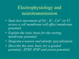
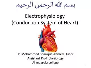
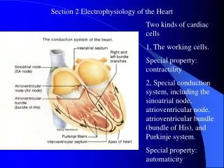
Cardiac Electrophysiology The properties which are especially important for understanding drug action on heart are: 1. Impulse generation Electrophysiologically, two types of myocardial fibres can be distinguished (a) Nonautomaticfibres:- These are the ordinary working myocardial fibres; cannot generate an impulse of their own. During diastole, the resting membrane potential remains stable (approximately 90 mv negative inside). When stimulated, they depolarize very rapidly (fast 0 phase) with considerable overshoot (+ 30 mv) → rapid return to near isoelectric level (phase- 1) → maintenance of membrane potential at this level for a considerable period (phase-2, plateau phase) during which Ca2+ ions flow in and bring about contraction → relatively rapid repolarization (phase 3) during which membrane Na+K+ pump gets activated and tends to restore ionic distribution to the resting pattern. Resting membrane potential, once attained, does not decay (stable phase-4). (b) Automatic fibres :- These are present in the sinoatrial (SA) and atrioventricular (A-V) nodes, and in the His-Purkinje system, i.e. specialized conducting tissue. In addition, patches of automatic tissue are present in the interatrial septum, A-V ring and around openings of the great veins. The most characteristic feature of these fibres is phase-4 or slow diastolic depolarization, i.e. after repolarizing to the maximum value, the membrane potential decays spontaneously. When it reaches a critical threshold value—sudden depolarization occurs automatically. Thus, they are capable of generating their own impulse. The rate of impulse generation by a particular fibre depends on the value of maximal diastolic potential, the slope of phase-4 depolarization and the value of threshold potential.
Transmembrane potential of automatic (Red) and nonautomatic (Purple) myocardial fibres recorded through intracellular electrodes. Normally, the SA node has the steepest phase-4 depolarization, undergoes self-excitation and propagates the impulse to the rest of the heart— acts as the pacemaker. Other fibres which are also undergoing phase-4 depolarization, but at a slower rate, receive the propagated impulse before reaching threshold value and remain as latent pacemakers.
2. Conduction: - • The rate of conduction through a fibre is a function of its membrane responsiveness, which is defined by rate of rise of AP (dv/dt) as a function of membrane potential at which activation occurs (Fig. VIII.3); a more completely polarized membrane depolarizes faster. This type of relationship is seen in atrial, ventricular and Purkinje fibres (fast channel fibres which depolarize by Na+ current), but not in SA and A-V nodal cells which remain refractory for some time even after attainment of maximal resting potential. • The Na+ channels get progressively inactivated as the resting membrane potential (RMP) drops over the –80 to –60 mV range. Consequently, less negative the RMP at which activation occurs, fewer are the Na+ channels available for activation—slope of ‘0’ phase depolarization, AP amplitude and conduction velocity are reduced. • A drug which reduces the slope of 0 phase (at any given resting membrane potential) will shift the membrane responsiveness curve to the right and impede conduction. The reverse occurs with a drug that shifts the curve to the left. Membrane responsiveness curve can also be altered by disease. • Small cells at the upper margin of A-V node have very low conduction velocity (20 mm/sec). Normally Purkinje fibres (PFs) have the highest conduction velocity (4000 mm/sec) except near their junction with the ventricular fibres ‘gate region’, or if they change over from fast channel to slow channel response. • Membraneresponsiveness curve of a myocardial fibre showing the relationship between membrane polarization and dv/dt of 0 phase. Note the depressant action of quinidine.
3. Excitability :- This property of a fibre is defined by the strength of stimulus required to elicit a response or to produce an AP. Hyperpolarization decreases excitability while small reductions in resting membrane potential increase excitability by respectively increasing and decreasing the gap between it and the threshold potential. Thus, in fast channel fibres excitability is generally super-normal during the end of phase-3. However, when the resting membrane potential is reduced to a value below the threshold potential, the fibre becomes inexcitable. 4. Refractory period :- Effective refractory period (ERP) which is the minimum interval between two propagating APs, is the most important. It is closely related to the AP duration (APD). An AP can be evoked in fast channel fibres even before complete repolarization, because Na+ channels recover in a voltage-dependent manner above the threshold potential. As such ERP/APD is <1. By contrast, the Ca2+ channels recover in a time-dependent manner progressively after the fibre has fully repolarized. Thus, in slow channel fibres ERP/ APD is > 1. Most antiarrhythmic drugs increase ERP/APD ratio.
Autonomic influences on cardiac electrophysiology and contractility on variables of cardiac function, because many cardiovascular drugs have indirect/secondary autonomic effects