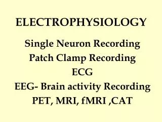
ELECTROPHYSIOLOGY
ELECTROPHYSIOLOGY. A subspecialty of cardiology. Nicole Harting P.A.-C. REFERENCES. CME Resources Inc. (board review coarse) Fogoros, Richard N., “Electrophysiologic Testing,” Fourth Edition. 2006. Heart Rhythm Society ( www.hrsonline.org ) Google Images. OBJECTIVES.

ELECTROPHYSIOLOGY
E N D
Presentation Transcript
ELECTROPHYSIOLOGY A subspecialty of cardiology Nicole Harting P.A.-C.
REFERENCES • CME Resources Inc. (board review coarse) • Fogoros, Richard N., “Electrophysiologic Testing,” Fourth Edition. 2006. • Heart Rhythm Society (www.hrsonline.org) • Google Images
OBJECTIVES • Overview of electrophysiology • Anatomy of the electrical system • Cardiac action potential • The heart rhythm • Arrhythmias • Atrial Fibrillation • Conduction system disease • Pacemakers • Cardiomyopathy and Internal Cardiac Defibrillators • Antiarrhythmics • Review of 12 lead EKGs and arrhythmias
OVERVIEW • The heart is an electrical organ. • The heart spontaneously generates electrical pulses by controlling the flux of calcium ions across the cardiac cell membrane. • These electrical impulses trigger muscle contraction. • The heart’s electrical impulses organize the sequence of muscle contractions during each heartbeat to optimize the cardiac stroke volume. • The pattern and timing of these impulses determine the heart rhythm. • Derangements in this rhythm can impair the heart’s ability to pump enough blood to meet the body’s demands.
ANATOMY OF THE ELECTRICAL SYSTEM • Sinoatrial node: located in the high right atrium. Initiates the “heart beat” (electrical impulse.) The impulse spreads radially across both atria. This impulse is represented by the P wave on an EKG. • Atrioventricular groove aka “skeleton” of the heart: The fibrous structure to which the valve rings are attached, and that separates the atria from the ventricles. Does notgenerate or spread electrical impulses, thus acts as an insulator. • Atrioventricular node (AV node): The beginning of the electrical connection between top and bottom of heart. The electrical impulse enters the node and the conduction (speed) is slowed down to allow atrial emptying and ventricular filling. This is represented by the PR interval on an EKG.
ANATOMY OF THE ELECTRICAL SYSTEM • His bundle: The most proximal part of the His-Purkinje system. The impulse is accepted from the AV node. The bundle penetrates the “skeleton” and delivers the impulse from the atrial to ventricular side of the heart. As the impulse spreads into the ventricular side of the heart it travels down the right and left bundle branches. • Purkinje fibers: The “end” of the conduction system. The fibers continue distally and outward to the furthermost reaches of the myocardium. • The impulse traveling down the entire His-Purkinje system is represented by the QRS complex on an EKG.
THE CARDIAC ACTION POTENTIAL • Cardiac cells are excitable. When they they are appropriately stimulated, tiny pores (channels) in the cell membrane open and close sequentially. This allows ions to travel back and forth leading to patterned changes in the transmembrane potential. • These changes graphed against time result in the cardiac action potential. The action potential is a reflection of the electrical activity of a single cardiac cell. • Three phases of the action potential: • 1) Depolarization: Rapid sodium channels in the membrane are stimulated to open. This is the “impulse” of the heart. Depolarization of one cell propogates the depolarization of the next cell… and so on until the impulse travels across the heart. The speed of depolarization determines the speed at which the impulse travels across the heart (heart rate). THUS, change the speed of depolarization and change the heart rate.
ACTION POTENTIAL CONTINUED • 2) Repolarization: Getting the ions back to where they started in order to depolarize again. It takes time to do this, and this time corresponds to the refractory period. During the refractory period, an electrical impulse cannot be generated. • 3) The resting phase: No net movement of ions across the cell membrane. • The action potential determines how quickly (conduction) an impulse can be generated and spread. It also determines how soon another impulse can be generated and spread. Overall, it determines heart rate. • Different areas/cells of the heart have different action potentials, thus different intrinsic rates and rhythms. • Arrhythmias result from an alteration of the action potential, NOT an “itch” or “irritation” in the heart. • Medications targeted at the heart rhythm affect the action potential of the cardiac cell. These medications change the shape of the action potential.
QUESTIONS TO DETERMINE HEART RHYTHM When looking at a 12 lead EKG it can be overwhelming. The BEST approach to reading an EKG is forming a system. Repeat the same steps every single time you look at a rhythm strip or EKG. “The Big 3”… the three main questions to determine heart rate and rhythm. • What are the atria doing? • What are the ventricles doing? • What is the relationship between the two? (don’t forget to check the heart rate during this step)
WHAT ARE THE ATRIA DOING? • First: look for P waves • Second: Are there P waves throughout the rhythm strip or EKG? • Third: Do the P waves march out? • Fourth: Analyze the P waves (shape, width) • All electrical impulses originating from the sinus node have the following: • P waves • Upright P waves in lead II • Downward P waves in lead aVR
WHAT ARE THE VENTRICLES DOING? • First: Are there QRS complexes? • Second: Are there QRS complexes throughout the entire rhythm strip or EKG? • Third: Do the QRS complexes march out? • Fourth: Analyze the QRS complex (shape, width) • If the heart rhythm is being “driven” by the atria then the QRS should be narrow. • If the ventricles are in control the QRS is ALWAYS wide. • Bundle branch blocks have wide QRS complexes.
WHAT IS THE RELATIONSHIP BETWEEN THE TWO? • Is every P wave followed by a QRS complex? • Is there a regular or irregular pattern? • What is the distance between the P wave and the QRS complex? Are the distances the same or different? • What is the rate? (normal, fast, slow) • If the QRS is wide… what does it look like in lead V1? (mostly up? mostly down? notched?) • Who is “driving” the rhythm? • Name the rhythm!
EKG • After answering “The Big 3” take closer look at the following… • What is the PR interval? • How wide is the QRS complex? • What does the ST segment look like? Is it flat, elevated or depressed? • What does the T wave look like? • What is the QT segment length? • Are there Q waves? • These will help answer: Is the patient having a heart attack? Is there ischemia? Is there a heart block? Is there QT prolongation?
SINUS RHYTHM (60-100 BPM) • Originating from the SA node. • Lead II the P wave is upright • Lead aVR the P is down • PR interval is < 0.2 seconds (200 ms) • every P is followed by a QRS
PREMATURE ATRIAL CONTRACTION • An early beat originating from the atria. • P wave is present • Narrow QRS • Many times the P wave morphology is different from sinus node beats.
SINUS BRADYCARDIA (<60 BPM) • Originating from the sinus node or ectopically in the atria • Rate <60bpm. • P waves are present
SINUS TACHYCARDIA (>100 BPM) • Originating from the sinus node. • Usually going between 100 and 130bpm. P waves should be present, upright in lead II and down in lead aVR. • Almost always a response to something such as a fever, excitement, infection, thyroid (hypo or hyper). Thus, almost always reversible.
ATRIAL TACHYCARDIA (130-150BPM) • Originating in the atria. • P waves present (unlike in supraventricular tachycardia- see next slide). • P waves may have a different morphology from sinus rhythm… aka: may not be up in lead II, down in aVR. May be “fatter”, notched, etc…
SUPRAVENTRICULAR TACHYCARDIA (>160BPM) • Originating above the ventricles… sinus node, atria or junction. • P waves may or may not be present • Narrow QRS complex • Supra= above • Ventricular= ventricles • Thus, a rhythm from above the ventricles.
MULTIFOCAL ATRIAL TACHYCARDIA • Originating from multipleregions of the atria, thus there are at least 3 different P wave morphologies and varying PR intervals. • Fast rate, usually between 100 and 120 at rest. • An irregular rhythm that is often confused with atrial fibrillation. • Very commonly associated with lung disease, often COPD. • If a patient has this rhythm and no known lung disease/issues they should be closely evaluated for it.
ATRIAL FLUTTER • Key words to remember for testssaw-tooth pattern. • Arrhythmia originating from the right atrium. • Typical atrial rate is 300 and ventricular rate around 150. • Can be a regular or irregular rhythm. • Ventricular rate can vary from slow, to ‘normal’ to fast.
ATRIAL FLUTTER • When diagnosing the rhythm it is important to mention the P wave to QRS complex ratio AND ventricular rate response… • “2:1 atrial flutter with rapid ventricular rates” … this means that there are 2 P waves for every QRS complex, and the overall heart rate is fast (like the rhythm strip on this slide) • “5:1 atrial flutter with slow ventricular response”… this means that there are 5 P waves for every QRS complex, and the overall heart rate is slow
ATRIAL FIBRILLATION • Most common sustained arrhythmia. • >2.6 million people in the United States have atrial fibrillation • 1 in 10 people >70 years of age have the arrhythmia. • Increasing frequency with increasing age. • Why we care so much…. According the New England Journal of Medicine, in a study released in 2010, 1 in 5 strokes are caused by atrial fibrillation. • An irregular rhythm originating from the left atrium. • Rate ranges from slow, to normal to fast. • A “cousin” of atrial flutter. Almost all patients with atrial flutter will have atrial fibrillation, and vice-versa. • No known cause of the arrhythmia, only some known correlations. • Commonly found in patients with hypertension, COPD, surgery pulmonary hypertension, atrial dilation, CHF, valvular disease.
ATRIAL FIBRILLATION • Three Types of a fib • Paroxysmal AF – Occurs sometimes and then stops. The AF may last for seconds, minutes, hours or days before the heart returns to its normal rhythm. People with this type of AF usually have more symptoms than others. As the heart goes in and out of AF, the pulse rate may change from slow to fast and back again in short periods of time. • Persistent AF – Persistent AF is when the AF does not stop by itself. Medications or a special type of electrical shock (called a cardioversion) is used to help the heart return to normal rhythm. If no treatment is given, the heart will stay in AF • Permanent AF – Permanent AF is when the AF cannot be fixed. Medications and controlled electrical shock cannot help return the heart to normal rhythm.
ATRIAL FIBRILLATION • Symptoms • Asymptomatic: usually in the elderly. Also common when the rate is ‘normal’ (between 60 and 100). • Palpitations: described as “racing heart,” “fluttering,” “butterflies in my chest” • Shortness of breath • Chest tightness or discomfort • Lightheadedness or dizziness (not vertigo) • Fatigue with “usual” activities… doing laundry, getting mail, getting dressed etc. • NOT associated with true syncope/LOC
ATRIAL FIBRILLATION • Effects of the arrhythmia on the body • Elevated Rates • Decreased cardiac output due to loss of atrial kick and decreased filling time of the ventricles (cause heart is dancing). • At all rates • Blood stasis in the left atrium increasing risk for left atrial appendage thrombus • Increased stroke risk due to above point
RHYTHM CONTROL APPROACH • Only if patient is able to be safely anticoagulated • Anticoagulate with IV Heparin and Coumadin bridging until INR therapeutic (>2.0) (if Pradaxa candidate then no need for bridging) – Remember this for year 2 • If asymptomatic and rate controlled: Coumadin x1 month then elective cardioversion. Initiate antiarrhythmic after cardioversion. • If symptomatic and/or difficult to rate control: cardiovert while on IV Heparin and initiate antiarrhythmic after. • Antiarrhythmic with a combination of rate control medication (BB, CCB, Digoxin) • Before cardioversion can be performed, evaluation for a thrombus needs to be completed. This is done with a transesophageal echocardiogram (TEE).
ANTICOAGULATION • Determine need for anticoagulation with CHADS 2 score (next slide). Moderate to high risk should be treated. • Determine if candidate. Patients should NOT be placed on Coumadin or Pradaxa if they have a history of massive GI bleed, recent bleeding in brain, unsteady gait, frequent falls. Basically…. Does the risk of bleeding outweigh the risk of stroke? • 1st line therapy: Coumadin or Pradaxa • 2nd line therapy: ASA 325mg and Plavix 75mg combination • 3rd line therapy: ASA 325mg
CHADS 2 SCORE - VASK • Stroke Risk Evaluation: CHADS 2 score • C: congestive heart failure 1 point • H: hypertension 1 point • A: age >_ 75 1 point • D: diabetes 1 point • S: stroke or TIA 2 points
“ABLATE AND PACE” • If a patient has recurrent, symptomatic atrial fibrillation with rapid rates an “ablate and pace” approach may be needed. • Implant a permanent pacemaker • Perform an AV nodal ablation (electrically disconnecting the atria from the ventricles.) • This resolves the symptoms, does NOT fix the arrhythmia
ATRIAL FIBRILLATION ABLATION In young patients we can attempt an atrial fibrillation ablation. It is a lengthy procedure (up to 6 hours) that cauterizes the tissue around the connection of the pulmonary veins to the left atrium. Atrial fibrillation often returns and a second ablation is (commonly) needed. • This is done in 2 ways: • Catheter ablation through the groin. Performed in the EP lab • MAZE procedure during an open heart procedure. Performed in OR.
KEY POINTS • Cardioversion: electrical shock delivered to the heart to return it to sinus rhythm • If the patient is hemodynamically unstable perform an emergent cardioversion. • Do NOT cardiovert someone (either electrically or with antiarrhythmics) if they are not properly anticoagulated. • Rhythm control vs rate control: NO difference in overall mortality. • Once you have atrial fibrillation it will come back. The longer you have it, the more frequently it will return and the longer it will last. • Electrically, the rhythm is NOT dangerous if rate controlled. We worry about the increased stroke risk. • If a patient is having recurrent strokes and no cause is found, monitor them with heart monitors as an outpatient to look for atrial fibrillation.
JUNCTIONAL RHYTHM (40-60 BPM) • Originating from the AV node (aka the junction). • P waves are either absent, down or after the QRS complex. • Often an “escape” rhythm… when the SA node and atria fail
ACCELERATED JUNCTIONAL (>60 BPM) • Originating from the AV node (junction) • Same exact rhythm as the previous slide… just faster!
VENTRICULAR RHYTHMS • Originate from the ventricles, thus the intrinsic rate is much slower than the atria. (Remember why the rate is slower?... different action potential!) • Wide QRS complex • NO P waves are present • These arrhythmias have a more grave prognosis and should be evaluated and treated promptly.
PREMATURE VENTRICULAR CONTRACTION • An early beat originating from the ventricles • No P wave • Wide QRS • Benign…
IDIOVENTRICULAR RHYTHM (20-40 BPM)ACCELERATED >40 BPM • Originating from the ventricles • No P waves present • Wide QRS • Slow rhythm
VENTRICULAR TACHYCARDIA (>100 bpm) • Originating from the ventricles. • No P wave and wide QRS • Fast rhythm… never, ever a good or benign rhythm
VENTRICULAR FIBRILLATION • Fatal rhythm originating in the ventricles • No discernable QRS complexes • Immediate defibrillation needed • *Bottom strip: ventricular tachycardia deteriorating into vfib*