Chapter 8 Visual System
Chapter 8 Visual System. Chris Rorden University of South Carolina Norman J. Arnold School of Public Health Department of Communication Sciences and Disorders University of South Carolina. Apparent motion. Visual Perception Events. Refraction of light rays by lens and cornea

Chapter 8 Visual System
E N D
Presentation Transcript
Chapter 8 Visual System • Chris Rorden University of South Carolina Norman J. Arnold School of Public Health Department of Communication Sciences and Disorders University of South Carolina
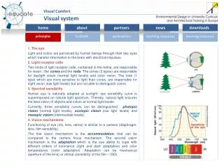
Visual Perception Events • Refraction of light rays by lens and cornea • Conversion of electromagnetic energy of light to nerve impulse • Transmission of action potential to CNS • Perception of visual image in visual cortices
Terminology • Optic Nerve • Visual fibers from retina to optic chiasm • Optic Tract • Optic fibers between chiasm to lateral geniculate body of thalamus or fibers that bypass thalamus to superior colliculus • Optic Radiation • Fibers project to visual cortex via geniculocalcarine fibers (optic radiation)
Visual Pathway • Cortical and subcortical processing
Visual Field • Visual Field: area you see before you - outside world • Retinal Field: Focused representation of visual field • Reversed (right/left, up/down) • Monocular Visual Field: Lateral portion perceived in only one eye • Binocular Visual Field: Common area seen by both eyes
Eyeball • Weighs 7.5 g and 2.4 cm long • 5/6 in orbital cavity • Anterior Chamber filled with aqueous humor • Made by choroid plexus of the ciliary processes • Drains through canal of Schlemm • Need to maintain pressure and link to circulatory system
Cavities and Chambers of Eyeball Anterior chamber Posterior chamber Anterior cavity Retina Choroid Sclera Posterior cavity Vitreous humor Fovea Optic disk Optic nerve Macula lutea
Ocular Layers • Fibrous Tunic (blue) • Sclera: White of eye • Cornea: Nonvascular and transparent fibrous region of eye • Vascular Tunic (yellow) • Choroid • Iris • Ciliary Body • Nervous Tunic (red) • Retina: Rods and Cones
Functions • Lens • Focuses images on the Retina • Ciliary Muscle • Regulates changes by lens (near and far vision) • Iris • Controls pupil size Aqueous humor Cornea Pupil Iris Lens Ciliary body Vitreous humor
Pupil, Iris, Scelera • Pupil • Iris • Scelera
Anatomy of Retina • Rods • Night vision • Cones • 3 types: sensitive to long, medium and short wavelength • Often red, green, blue but actual peak sensitivity is yellow, yellowish-green, and blue • Bipolar Cells • Ganglion Cells • Light passes through cell layers and then back to the ganglion cells.
Photo receptors http://www.webexhibits.org/colorart/ganglion.html http://web.mit.edu/bcs/schillerlab/research/A-Vision/A3-1.html
Blindspot • There are no rods or cones in the optic disk. • Close your right eye, and look at the 'x' in the figure. Move either closer or further away from the screen until you notice the that circle with the dot inside vanishes altogether.
Photosensors • Cones (30 million) • Discriminate color and sharp vision • Cone cells in macula lutea • fovea centralis • Rods (100 million) • Discriminate in dim light • Sensitive to shape and movement • Lateral peripheral retina • You can often see things better at night if you do not look directly at them! • We will not cover photochemistry of retina and optical mechanism.
Central Visual Mechanism • Visual pathway from retina to primary visual cortex • Optic nerve fibers exit optic foramina and move to optic chiasm • Optic tract move to lateral geniculate body (Remember it is part of thalamus) • Travel to occipital lobe to visual cortex
Visual Pathway • Each eye sees both left and right visual field. • Ipsilateral information crosses over at optic chiasm. • Some connections to superior colliculi. • Reflexive eye movments • Others go to thalamus (lateral geniculate nuclei) and then cortex.
Retinal Representation • Nasal and temporal visual fields • When you are looking at an object, these form the medial (nasal) and lateral (temporal) hemifield of vision for each eye. • Reversed to opposite halves of retinal representative fields • Also inverted • Nasal visual fields project to temporal retinal fields and do not cross at optic chiasm • Temporal visual field project to nasal retinal fields and cross at optic chiasm
Lateral Geniculate Nucleus to Visual Cortex • Optic Radiation (geniculocalcarine fibers; Meyer’s Loop) runs under temporal lobe to occipital lobe Lateral Geniculate Nucleus (Thalamus) V1 Primary Visual Cortex (BA17)
Reflexes • Pupillary Light Reflex • Involves Edinger-Westphal Nucleus and oculomotor CN (III) • Pupil contracts with light (consensual response) • Damage to system may be due to Horner’s syndrome (always constricted pupil) or CN III lesion damage to afferents to one eye • Accommodation Reflex: The focus reflex • Modifies lens curvature when object moves closer to eyes • Lens flexibility important • Lens tends to become less flexible around age 45
Horner’s syndrome • Injury to sympathetic nervous system • First-order neuron disorder: Central lesions that involve the hypothalamospinal pathway (e.g. transection of the cervical spinal cord). • Second-order neuron disorder: Preganglionic lesions (e.g. compression of the sympathetic chain by a lung tumor). • Third-order neuron disorder: Postganglionic lesions at the level of the internal carotid artery (e.g. a tumor in the cavernous sinus). • ptosis (drooping eyelid), miosis (constricted pupil) and dilation lag.
Clinical Conditions • Hypermetropia (farsightedness) • Can see distant objects normally but problem in near objects • Due to short eyeball and inadequate refractory power of the lens • Myopia (nearsightedness) • Can see near objects but not distant • Due to abnormally long eyeball and too strong refractory power
Clinical Conditions Standard Cylindrical • Astigmatism • Focus disorder of vertical and horizontal rays • Caused by irregular shape or the cornea, lens, or both • Can typically be corrected with glasses with relatively cylindrical rather than dish shaped lenses.
Clinical conditions • Color vision disorders (usually males) • First documented by John Dalton (1798) • Dichromacy: Loss single type of cone, e.g. of long (yellow, protanopia), medium (yellow-green, deuteranopia) or short (blue, tritanopia) wavelength. • Monochromacy: Total color blindness due to absence of cones or abnormal cones Normal Protanopia Deuteranopia Tritanopia
Other Common Disorders • Presbyopia - decrease in vision with age • Cataract - Increase in protein in lens • Glaucoma - Increased intraocular pressure • Infections - Inflammation of the eye • Retinitis Pigmentosa - familial disorder causing loss of rod cells. Includes peripheral visual loss and night blindness
Visual defects following stroke Neglect • Damage to early visual centers causes blindness (see next slides). • Damage to temporal/parietal lobes cause: • Neglect: failure to respond to contralesional stimuli (usually right hemisphere injury) • Achormatopsia: color blindness • Akinetopsia: Motion blindness (very rare) • Agnosia: failure to recognize objects • Ataxia: reaching deficits • Simultanagnosia: only see one thing at a time Agnosia: can copy But not recognize
V1 (BA 17) • Primary visual cortex (V1) lies in calcarine fissure. • Complete damage leads to Homonymous hemianopia. • Partial damage leads to scotomas • Point-to-point mapping with retina.
Types of Field Defects • Left optic tract carries info from right visual field in each eye • Right optic tract carries info from left visual field in each eye • Simplified in that some overlapping present
Types of Field Defects L R A Monocular blindness A B Bitemporal hemianopsia B C C Nasal hemianopsia D E D Homonymous hemianopia F E Homonymous left Superior quadrantopsia F Homonymous left Inferior quadrantopsia
Visual Field Defects • Homonymous • Similar regions affected in each eye • i.e. Right visual fields of both eyes • Heteronymous • Different regions affected in each eye • i.e. Left visual field of one eye and right visual field of other eye
Specific Deficits • Monocular Blindness: Blindness in one eye due to optic nerve lesion before optic chiasm • Bitemporal (Heteronymous) Hemianopsia: Loss of temporal visual fields of each eye, lesion at optic chiasm • Nasal Hemianopsia: Loss of nasal vision in one eye due to lesion in lateral edge of optic chiasm. • Homonymous Hemianopsia: Loss of left or right visual fields for both eyes due to lesion in right optic tract • Upper Left Quadrantanopsia: Loss of vision in left upper quadrant of each eye due to lesion in Meyer’s Loop • Lower left Quadrantanopsia: Loss of vision in lower left quadrant of each eye due to lesion in medial fibers of visual tract