Download
1 / 30
1.93k likes | 9.48k Vues
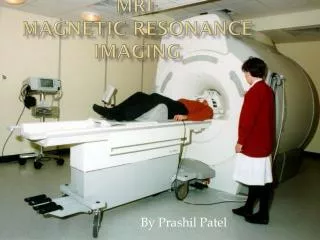
Magnetic Resonance Imaging (MRI). Introduction The Components The Technology Physics behind MR Conclusion Presented by Salwa Aziz & Christina Derbidge. Introduction. What is MRI?

E N D
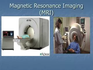
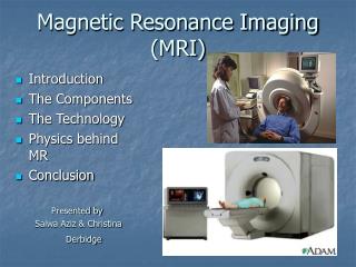
Magnetic Resonance Imaging (MRI) • Introduction • The Components • The Technology • Physics behind MR • Conclusion Presented by Salwa Aziz & Christina Derbidge
Introduction • What is MRI? • Magnetic resonance imaging (MRI) is a spectroscopic imaging technique used in medical settings to produce images of the inside of the human body. • MRI is based on the principles of nuclear magnetic resonance (NMR), which is a spectroscopic technique used to obtain microscopic chemical and physical data about molecules • In 1977 the first MRI exam was performed on a human being. It took 5 hours to produce one image.
Introduction • How Does it Work? • The magnetic resonance imaging is accomplished through the absorption and emission of energy of the radio frequency (RF) range of the electromagnetic spectrum.
The Components: • A magnet which produces a very powerful uniform magnetic field. • Gradient Magnets which are much lower in strength. • Equipment to transmit radio frequency (RF). • A very powerful computer system, which translates the signals transmitted by the coils.
The Magnet • The most important component of the MRI scanner is the magnet: • The magnets currently used in scanners today are in the .5-tesla to 2.0-tesla range (5,000 to 20,000-gauss). Higher values are used for research. • Earth magnetic field: 0.5-gauss
The Magnet (cont.) • There are three types of magnets used in MRI systems: • Resistive magnets • Permanent magnets • Super conducting magnets (the most commonly used type in MRI scanners). • In addition to the main magnet, the MRI machine also contains three gradient magnets. These magnets have a much lower magnetic field and are used to create a variable field.
The Technology • How Does It All Work? • Spin: • The atoms that compose the human body have a property known as spin(a fundamental property of all atoms in nature like mass or charge). • Spin can be thought of as a small magnetic field and can be given a + or – sign and a mathematical value of multiples of ½. • Components of an atom such as protons, electrons and neutrons all have spin.
The Technology (cont.) • Spin(cont.): • Protons and neutron spins are known as nuclear spins. • An unpaired component has a spin of ½ and two particles with opposite spins cancel one another. • In NMR it is the unpaired nuclear spins that produce a signal in a magnetic field.
The Technology (cont.) • Human body is mainly composed of fat and water, which makes the human body composed of about 63% hydrogen. • Why Are Protons Important to MRI? • positively charged • spin about a central axis • a moving (spinning) charge creates a magnetic field. • the straight arrow (vector) indicates the direction of the magnetic field.
The Technology (cont.) • When placed in a large magnetic field, hydrogen atoms have a strong tendency to align in the direction of the magnetic filed • Inside the bore of the scanner, the magnetic field runs down the center of the tube in which the patient is placed, so the hydrogen protons will line up in either the direction of the feet or the head. • The majority will cancel each other, but the net number of protons is sufficient to produce an image.
The Technology (cont.) • Energy Absorption: • The MRI machine applies radio frequency (RF) pulse that is specific to hydrogen. • The RF pulses are applied through a coil that is specific to the part of the body being scanned.
The Technology (Cont.) Resonance (cont.) The gradient magnets are rapidly turned on and off which alters the main magnetic field. • The pulse directed to a specific area of the body causes the protons to absorb energy and spin in different direction, which is known as resonance Frequency (Hz) of energyabsorption depends on strength of external magnetic field.
The Technology (cont.) • The resonance frequency, 0, is referred to as the Larmor frequency.
The Technology (cont.) • Imaging: • When the RF pulse is turned off the hydrogen protons slowly return to their natural alignment within the magnetic field and release their excess stored energy. This is known as relaxation. • What happens to the released energy? • Released as heat OR • Exchanged and absorbed by other protons OR • Released as Radio Waves.
The Technology (cont.) • Measuring the MR Signal: • the moving proton vector induces a signal in the RF antenna • The signal is picked up by a coil and sent to the computer system. the received signal is sinusoidal in nature • The computer receives mathematical data, which is converted through the use of a Fourier transform into an image.
Physics of MRIIt is an interplay of • Magnetism • Resonance
Fig: 1. A) The top spinning in the earth's gravity. The gravity tries to pull it down but it stays upright due to its fast rotation. B) A charge spinning in the magnetic field Bo.
Fig: 2. A) The protons spinning in the nature, without an external strong field. The directions of spins are random and cancel out each other. The net magnetization is nearly 0. B) In the presence of a large external magnetic field Bo the spins align themselves either against (low energy state) or along (low energy state). There is a slight abundance of spins aligned in the low energy state.
Fig: 3. A) The compass needle (a small magnet) aligns itself with a N/S-S/N direction when placed in a large magnetic field. B) When another strong magnet is brought near the aligned compass needle the magnetic fields of all three magnets interact in such a way that the mobile, weakest magnet (the compass needle) realigns itself away from its original orientation. C) When the perturbing magnetic field is removed suddenly the compass needle magnet realigns itself with the large external magnet field, but before realigning, it wobbles around the point of stability and gradually comes to rest.
Fig: 4. The spin of a proton can be represented by a vector B with a direction and magnitude. Its relation to the direction of the external magnetic field Bo is represented by an angle.
Fig: 5. A) The spin of a proton aligned to Bo in the Z-axis. B) An external perturbing magnetic field, B1, is applied which knocks the vector out of its axis, which now is aligned at a new angle with respect to Bo. C) As the perturbing field B1 is removed the vector gradually starts returning back to its original state and D) begins to wobble
Fig: 6. A) The falling water rotates a wheel to which a magnet is attached. When this magnet rotates it induces an alternating current in a coil of wire which can be detected. B) A magnetic field (spin of a proton) rotating near a coil of MR antenna induces a similar current in the loop which can be detected.
Resonance • Fig: 7. The gradient coils. A) the body placed in the core of the magnet with B0 aligned to its long axis. B) the gradient coil oriented in the Z-axis (along the long axis of the body) which gradually and linearly increases from left to right. C) At the center of the gradient field, the frequency is equal to that of B0, but at a distance x the field changes by a factor of B0.
Resonance • The resonance equation shows that the resonance frequency n of a spin is proportional to the magnetic field, Bo, it is experiencing. n = g Bo • Where g is the gyromagnetic ratio. [the ratio of the magnetic moment of a spinning charged particle to its angular momentum]
Recap: What Does the Image Represent? • For every unit volume of tissue, there is a number of cells, these cells contain water molecules, each water molecule contain one oxygen and two hydrogen atoms. • Each hydrogen atom contains one proton in its nucleus. Different tissues thus produce different images based on the amount of their hydrogen atoms producing a signal
Why MRI ? • Utilizes non ionizing radiation. (unlike x-rays). • Ability to image in any plane. (unlike CT scans). • Very low incidents of side effects. • Ability to diagnose, visualize, and evaluate various illnesses. The only better way to see the insides of your body is to cut you open!
References • Ballinger, Ray. Basics of MRI. 1994-1996. http://www.mritutor.org/mritutor/basics.htm Retrieved: 7/7/03 • Buckwalter, Ken, M.D. Magnetic Resonance Imaging. http://www.indyrad.iupui.edu/public/lectures/mri/iu_lectures/mri_homepage.htm Retrieved: 7/6/2003 • Gould, Todd, RT, MR, ARRT. How MRI Works. http://electronics.howstuffworks.com/mri7.htm Retrieved:7/5/2003 • Hornak, Joseph, PhD. The Basics of MRI. 1996-2003. http://www.cis.rit.edu/htbooks/mri/index.html • Nagasaki School of Medicine, Department of Radiology. Basics of MRI-I. http://www.med.nagasakiu.ac.jp/radiolgy/MRI%20of%20the%20FOOT/MRI-CDNUH/nf-basic1.html Retrieved 7/7/03.