
SlideServe
Menu
Préférences
© SlideServe · v2026

Magnetic Resonance Imaging (MRI)

suchi avatar
19 diapositives · 1.1K Views · Mar 18, 2014

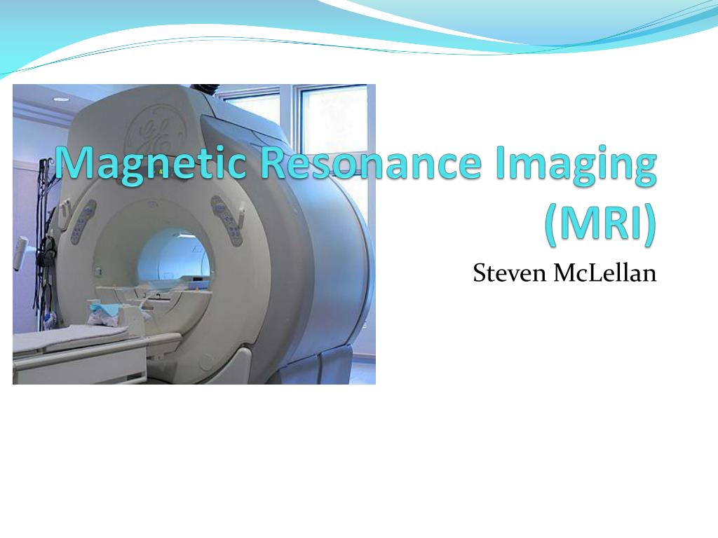
Magnetic Resonance Imaging (MRI). Steven McLellan. What is MRI?. Produces very clear, detailed pictures of the organs and structures in the body It is a form of medical imaging that uses no Ionizing radiation
Magnetic Resonance Imaging (MRI) - slide 1
01 / 19Magnetic Resonance Imaging (MRI)
1 / 17

Télécharger la présentation

Magnetic Resonance Imaging (MRI)
Une image / un lien ci-dessous est fourni (tel quel) pour télécharger la présentation

Politique de téléchargement : le contenu du site vous est fourni TEL QUEL pour votre information et votre usage personnel ; il ne peut être vendu, concédé sous licence ni partagé sur d’autres sites sans l’accord de son auteur.Le contenu vous est fourni TEL QUEL, pour votre information et votre usage personnel uniquement. Téléchargez la présentation en cliquant sur ce lien. Lors du téléchargement, si vous ne parvenez pas à télécharger une présentation, l’éditeur a peut-être supprimé le fichier de son serveur.Pendant le téléchargement, si vous ne parvenez pas à obtenir une présentation, le fichier a peut-être été supprimé par l’éditeur.

F I N

Plus de contenu associé

Transcription de la présentation
  1. Magnetic Resonance Imaging(MRI) Steven McLellan

  2. What is MRI? • Produces very clear, detailed pictures of the organs and structures in the body • It is a form of medical imaging that uses no Ionizing radiation • MRI makes use of the property of Nuclear magnetic resonance (NMR) to image nuclei of atoms inside the body.

  3. History • The first MR image was published in 1973 • The first studies performed on humans were published in 1977 • Created by Dr. Raymond V. Damadian, Dr. Larry Minkoff and Dr. Michael Goldsmith • In 2003, The 2003 Nobel Prize in Physiology or Medicine was awarded to Paul C Lauterbur and Peter Mansfield • Made new MR imaging techniques • Faster and more efficient

  4. Common Uses • Physicians use the MR examination to help diagnose or monitor treatment for conditions such as: • Tumors and other cancer related abnormalities. • Certain types of heart problems. • Blockages or enlargements of blood vessels • Diseases of the liver, such as cirrhosis, and that of other abdominal organs. • Diseases of the small intestine, colon, and rectum

  5. Common Uses

  6. How does it work? • An MRI machine uses a powerful magnetic field to align the magnetization of some atoms in the body. • radio frequency fields systematically alter the alignment of this magnetization • This causes the nuclei to produce a rotating magnetic field detectable by the scanner • This information is recorded to construct an image of the body.

  7. How does it work?

  8. How does it work? • Images are constructed when protons in different tissues return to equilibrium state at different rates. • Five variables effect these rates • Spin Density: Concentration of nuclei in tissue processing in a given region under a magnetic field. • T1: Longitudinal relaxation time • T2: Transverse relaxation time • Flow: Shows blood flow, CSF flow • Spectral Shifts: Angle/zoom the picture is taken from.

  9. Basic MRI Scans • T1-weighted: Differentiate fat from water • Water is Darker, fat is brighter • Provide good gray matter/white matter contrast in brain. • T2-weighted: Differentiate fat from water • Fat shows darker, and water lighter. • Good for imaging edema • Abnormal accumulation of fluid beneath the skin or in one or more cavities of the body

  10. Common Uses

  11. Common Uses

  12. Specialized MRI Scans Diffusion MRI • Measures diffusion of water through biological tissues. • Diffusion may be anisotropic (unequal physical properties along different axes) • Diffusion Tensor Imaging (DTI) • Examine areas of neural degeneration and demyelination in diseases such as Multiple Sclerosis (MS)

  13. Diffusion MRI

  14. Specialized MRI Scans Magnetic resonance angiography (MRA) • Generates pictures of arteries. • Evaluates the arteries of the neck and brain, the thoracic and abdominal aorta, the renal arteries, and the legs • Uses gadolinium injection as paramagnetic contrast agent • Magnetic resonance venography (MRV) is a similar procedure that is used to image veins.

  15. Magnetic Resonance Angiography

  16. Safety Risks • MRI’s create up to 120dB • Equivalent to jet engine at take off. • Contraindications: • Pacemakers, Vagus Nerve Stimulators, implantable defibrillators, insulin pumps, deep brain stimulators • Any electronic or magnetized foreign bodies (surgical prosthesis) • Peripheral nerve stimulation (PNS) • Rapid switching on and off of the magnetic field gradients is capable of causing nerve stimulation

  17. During Procedure • People hold the part of their body being scanned motionless for 30-60 minutes. • Procedure is done in multiple parts. • Takes time to switch between different scans and fields of view.

  18. Future? • More detailed images • All MRIs use color? • Better pictures of bone structures • Shift from x-rays and CT scans to MRI • New Scanning sequences

  19. Citations • Dyson, Sue J. Magnetic Resonance Imaging. Philadelphia, PA: Elsevier, 2007. Print. • Hashemi, Ray H., William G. Bradley, and Christopher J. Lisanti. MRI: the Basics. Philadelphia, PA: Lippincott Williams & Wilkins, 2010. Print. • "Magnetic Resonance Imaging (MRI) - Body." RadiologyInfo - The Radiology I nformation Resource for Patients. Radiological Society of North America, Inc., 15 Mar. 2010. Web. 06 Mar. 2011. <http://www.radiologyinfo.org/en/info.cfm?pg=bodymr>. • Radiology". http://radiology.rsna.org/content/204/1/272.long. Retrieved 2 August 2010. • Westbrook, Catherine. MRI. John Wiley & Sons, Incorporated, 2009. Print.