
V5: Cell differentiation
V5: Cell differentiation. Complex genomes can generate a range of different cell types in a highly ordered and reproduceable manner. Epigenetic modifications are important for ‘programming’ lineage determination and cellular identity during development.

V5: Cell differentiation
E N D
Presentation Transcript
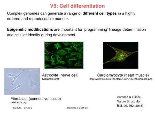
V5: Cell differentiation Complex genomes can generate a range of different cell types in a highly ordered and reproduceable manner. Epigenetic modifications are important for ‘programming’ lineage determination and cellular identity during development. Astrocyte (nerve cell) Cardiomyocyte (heart muscle) (wikipedia.org) (http://www.kcl.ac.uk/content/1/c6/01/66/46/gautel3.jpeg Cantone & Fisher, Nature Struct Mol Biol. 20, 292 (2013) Fibroblast (connective tissue) (wikipedia.org) Modeling of Cell Fate
Epigenetic stability • In somatic tissues, CpG islands at housekeeping or developmental promoters are largely unmethylated, whereas non-regulatory CpGs distributed elsewhere in the genome are largely methylated. • This DNA methylation landscape is relatively static across all somatic tissues. • Most of methylated CpGs are pre-established and inherited through cell division.How? • In at least two phases of the life cycle of mammals, epigenetic stability is globally perturbed: • when gametes fuse to form the zygote and • when gamete precursors (primordial germ cells; PGCs) develop and migrate in the embryo. • This in vivo ‘reprogramming’ of the epigenetic landscape signals the reacquisition of totipotency in the zygote and the formation of the next generation through PGCs. Cantone & Fisher, Nature Struct Mol Biol. 20, 292 (2013) Modeling of Cell Fate
Waddington: Epignetic landscape Conrad H. Waddington 1956: "Principles of Embryology“; www.nature.com Konrad Hochedlinger and Kathrin Plath, Development 136, 509-523 (2009) Modeling of Cell Fate
Zygotes - fertilization In living organisms that reproduce sexually, development starts from a single cell, the zygote (dt: befruchtete Eizelle). Zygotes are usually produced by a fertilization event between two haploid cells — an ovum from a female and a sperm cell from a male—which combine to form the single diploid cell. Human sperm and egg (sex cells) have one complete set of chromosomes from the male or female parent. Sex cells, also called gametes, combine to produce somatic cells. Somatic cells therefore have twice as many chromosomes. The haploidity number (n=23 in humans) is the number of chromosomes in a gamete. A somatic cell has twice that many chromosomes (2n=46). www.wikipedia.org Modeling of Cell Fate
some terms from developmental biology somatic cells = cells forming the body of an organism germ cells (dt. Keimzelle, Ovolum) are part of the germline. germline (dt. Keimbahn)= line of germ cells that have genetic material that may be passed to a child/embryo. Germline cells are immortal. Gametocyte = eukaryotic germ cell; includes spermatocytes (male) and oocytes (female) primordial germ cells : predecessors of germ cells. They migrate to the gonadal ridge (precursor of gonads). They may be detected from expression of Stella (gene) gonad (dt. Keimdrüse) www.wikipedia.org Modeling of Cell Fate
Germ line development Germline cells are produced by embryonic cleavage. Cleavage: division of cells in the early embryo. The zygotes of many species undergo rapid cell cycles with no significant growth. The different cells derived from cleavage are called blastomeres and form a compact mass called the morula (because it resembles a mulberry/ dt. Maulbeere). Cleavage ends with the formation of the blastula. Cleavage in mammals is slow. Cell division takes 12 – 24 hours and is asynchronous. www.wikipedia.org Modeling of Cell Fate
From left to right, the morula-stage mouse embryo (embryonic day 2.5; E2.5) holds a core of pre-ICM (inner cell mass) cells that turn into ICM cells at cavitation/ blastulation (E3–E4). At this stage, embryonic stem cell (ESC) and Trophoblast Stem Cell (TSC) cell lines can be derived. As the blastocyst fully expands, the ICM delaminates giving rise to a primitive ectoderm and a primitive endoderm layer. The blastocyst's outer cells are termed trophectoderm. In mammals, the ICM will ultimately form the "embryo proper", while the trophectoderm will form the placenta and other extra-embryonic tissues.[ At E6 and subsequently, the embryo will start gastrulating. This process involves the formation of a mesoderm layer between ectoderm and endoderm, and the formation of the primordial germ cells (PGCs). The pluripotent cells of the embryo are tracked in green. Boiani & Schöler, Nat Rev Mol Cell Biol 6, 872 (2005) Modeling of Cell Fate
3 primary germ cell layers The ectoderm is the outer layer of the early embryo. It emerges first and forms from the outer layer of germ cells. The ectoderm differentiates to form the nervous system (spine, peripheral nerves and brain), tooth enamel and the epidermis. It also forms the lining of mouth, anus, nostrils, sweat glands, hair and nails. The endoderm develops at the inner layer. Its cells differentiate to form the gastrointestinal tract, the respiratory tract, endocrine glands and organs, auditory systems, and the urinary system. The mesoderm is the middle layer. It differentiates to give rise to a number of tissues and structures including bone, cartilage (dt: Knorpel), muscle, connective tissue (including that of the dermis), the middle layer of the skin, blood vascular, reproductive, excretory and urinogenital systems and contributes to some glands. www.wikipedia.org Modeling of Cell Fate
Developmental Glossary (I) Inner cell mass (ICM): Cells ofthe blastocyst embryo that appear transiently during developmentand give rise to the three germ layers of the developing embryo. Embryonicstem (ES) cells: Pluripotent cell line derived from the ICM uponexplantation in culture. In vitro, ES cells can differentiate intomany different lineages and cell types. Upon injectioninto blastocysts, ES cells can give rise to all tissues including thegermline. Primordial germ cells (PGCs): In vivo, PGCs give rise to oocytesand sperm. When explanted in vitro, PGCs give rise to embryonic germ (EG) cells. Hochedlinger, Development 136, 509 (2009) Modeling of Cell Fate
Adult stem cells Embryonic stem cells only exist in the early embryo. We all possess adult stem cells, from which new specialized cells are formed throughout our life time. Adult cells exist predominantly in bone marrow (dt. Knochenmark), but also in skin, fat tissue, umbilical cord (dt. Nabelschnur), brain, liver, and in pancreas (dt. Bauchspeicheldrüse). Adult cells in cell culture have a much reduced ability of self regeneration and a reduced ability for differentiation compared to embryonic stem cells. For example, neural stem cells can differentiate to all cell types of neural tissue (neorons, glia), but likely not into liver or muscle cells. www.wikipedia.org Modeling of Cell Fate
Epigenetic programming and reprogramming during the mouse life cycle. Two populations of pluripotent cells can be established ex vivo within the time window in which extensive epigenetic reprogramming takes place. These cells are ESCs and embryonic germ cells (EGCs) that are derived from the inner cell mass of the blastocyst and from the PGCs at E8.5–E13.5, respectively. Major remodeling events (e.g. DNA demethylation and X-chromosome reactivation) are highlighted in the figure by colored arrows. TE, trophoectoderm; PE primitive endoderm. Cantone & Fisher, Nature Struct Mol Biol. 20, 292 (2013) Modeling of Cell Fate
Haematopoiesis Haematopoiesis (from Ancient Greek: αἷμα, "blood"; ποιεῖν "to make") is the formation of blood cellular components. All cellular blood compo-nents are derived from haematopoietic stem cells. In a healthy adult person, approximately 1011–1012 new blood cells are produced daily in order to maintain steady state levels in the peripheral circulation. Development of different blood cells from haematopoietic stem cell to mature cells www.wikipedia.org Modeling of Cell Fate
Differentiation Zygotes contain DNA derived from both the mother and the father, and this provides all the genetic information necessary to form a new individual. This property is named „totipotency“ (latin: totus – all, potentia – power/ability). Continuous cell division produces daughter cells that start to specialize on individual functions. This developmental process of cells and tissue from a less specialized to a more specialized state is called differentiation in developmental biology. www.wikipedia.org Modeling of Cell Fate
Glossary I Totipotency Ability of a cell to giverise to all cells of an organism, including embryonic and extraembryonictissues. Zygotes are totipotent. Pluripotency Ability of a cellto give rise to all cells of the embryo. Cells of the innercell mass (ICM) and its derivative, embryonic stem(ES) cells, are pluripotent. Multipotency Ability of a cellto give rise to different cell types of a given cell lineage.These cells include most adult stem cells, such as gut stemcells, skin stem cells, hematopoietic stem cells and neuralstem cells. Unipotency Capacity of a cell to sustain only onecell type or cell lineage. Examples are terminally differentiatedcells, certain adult stem cells (testis stem cells) and committedprogenitors (erythroblasts). Hochedlinger, Development 136, 509 (2009) Modeling of Cell Fate
Epigenetic changes during in vivo reprogramming Global DNA and histone modifi-cations that lead to transcriptional activation of the embryonic genome between the late zygote (paternal genome only) and the 2-cell stage. Protamines are small, arginine-rich, nuclear proteins that replace histones late in the haploid phase of spermatogenesis and are believed essential for sperm head conden-sation and DNA stabilization. In humans, 10-15% of the sperm's genome is packaged by histones thought to bind genes that are essential for early embryonic development (www.wikipedia.org). Gamete genomes undergo different epigenetic programs after fertilization. The paternal genome is mostly subject to epigenetic remodeling at the zygote stage. The maternal genome gradually loses repressive modifications during the subsequent cleavage divisions. Cantone & Fisher, Nature Struct Mol Biol. 20, 292 (2013) Modeling of Cell Fate
Epigenetic changes during germline development Global epigenetic changes during germline development from PGC specification (E6.5) to the mitotic/meiotic arrest at E13.5. Two major reprogramming phases can be distinguished during PGC migration toward the genital ridges (E7.5–E10.5) and upon their arrival into the gonads (E10.5–E12.5). Cantone & Fisher, Nature Struct Mol Biol. 20, 292 (2013) Modeling of Cell Fate
Summary Epigenetic remodelling is responsible for cellular differentiation. Altering chromatin structure will affect accessibility of genes and, hence, alter the transcriptional program in cells. Open question: - which genes/proteins are the drivers/master regulators? - Does epigenetics regulate transcription, or does transcription regulate epigenetics, or are both closely interlinked? - How can one study such combined epigenetic + gene-regulatory networks by computational modeling? www.wikipedia.org Modeling of Cell Fate


