Download
1 / 5
50 likes | 104 Vues
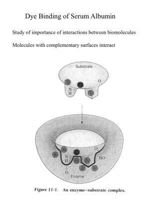
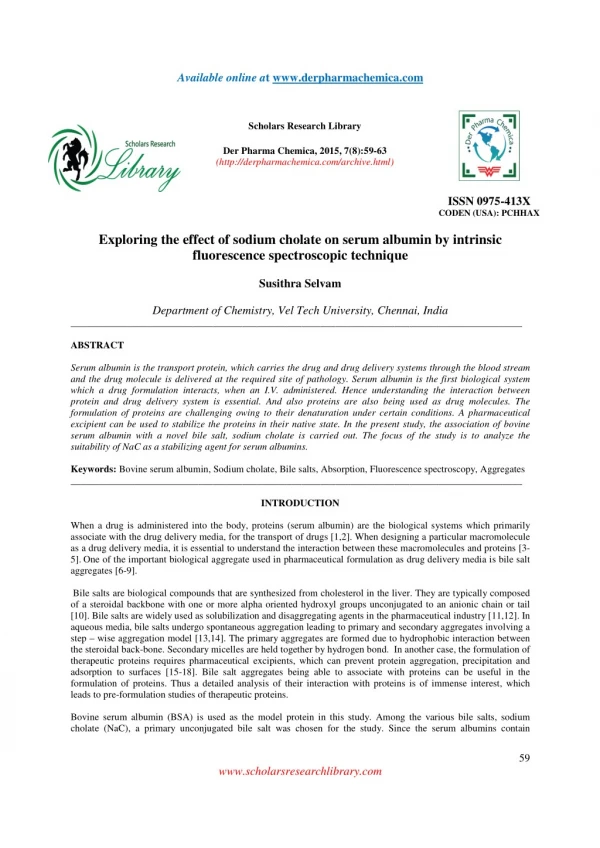
Serum albumin is the transport protein, which carries the drug and drug delivery systems through the blood stream<br>and the drug molecule is delivered at the required site of pathology. Serum albumin is the first biological system<br>which a drug formulation interacts, when an I.V. administered. Hence understanding the interaction between<br>protein and drug delivery system is essential. And also proteins are also being used as drug molecules. The<br>formulation of proteins are challenging owing to their denaturation under certain conditions. A pharmaceutical<br>excipient can be used to stabilize the proteins in their native state. In the present study, the association of bovine<br>serum albumin with a novel bile salt, sodium cholate is carried out. The focus of the study is to analyze the<br>suitability of NaC as a stabilizing agent for serum albumins. <br>

E N D
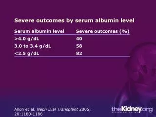
ailable online a Av www.derpharmachemica.com t Scholars Research Library Der Pharma Chemica, 2015, 7(8):59-63 (http://derpharmachemica.com/archive.html) ISSN 0975-413X CODEN (USA): PCHHAX Exploring the effect of sodium cholate on serum albumin by intrinsic fluorescence spectroscopic technique Susithra Selvam Department of Chemistry, Vel Tech University, Chennai, India ____________________________________________________________________________________________ ABSTRACT Serum albumin is the transport protein, which carries the drug and drug delivery systems through the blood stream and the drug molecule is delivered at the required site of pathology. Serum albumin is the first biological system which a drug formulation interacts, when an I.V. administered. Hence understanding the interaction between protein and drug delivery system is essential. And also proteins are also being used as drug molecules. The formulation of proteins are challenging owing to their denaturation under certain conditions. A pharmaceutical excipient can be used to stabilize the proteins in their native state. In the present study, the association of bovine serum albumin with a novel bile salt, sodium cholate is carried out. The focus of the study is to analyze the suitability of NaC as a stabilizing agent for serum albumins. Keywords: Bovine serum albumin, Sodium cholate, Bile salts, Absorption, Fluorescence spectroscopy, Aggregates ____________________________________________________________________________________________ INTRODUCTION When a drug is administered into the body, proteins (serum albumin) are the biological systems which primarily associate with the drug delivery media, for the transport of drugs [1,2]. When designing a particular macromolecule as a drug delivery media, it is essential to understand the interaction between these macromolecules and proteins [3- 5].One of the important biological aggregate used in pharmaceutical formulation as drug delivery media is bile salt aggregates [6-9]. Bile salts are biological compounds that are synthesized from cholesterol in the liver. They are typically composed of a steroidal backbone with one or more alpha oriented hydroxyl groups unconjugated to an anionic chain or tail [10]. Bile salts are widely used as solubilization and disaggregating agents in the pharmaceutical industry [11,12]. In aqueous media, bile salts undergo spontaneous aggregation leading to primary and secondary aggregates involving a step – wise aggregation model [13,14]. The primary aggregates are formed due to hydrophobic interaction between the steroidal back-bone. Secondary micelles are held together by hydrogen bond. In another case, the formulation of therapeutic proteins requires pharmaceutical excipients, which can prevent protein aggregation, precipitation and adsorption to surfaces [15-18]. Bile salt aggregates being able to associate with proteins can be useful in the formulation of proteins. Thus a detailed analysis of their interaction with proteins is of immense interest, which leads to pre-formulation studies of therapeutic proteins. Bovine serum albumin (BSA) is used as the model protein in this study. Among the various bile salts, sodium cholate (NaC), a primary unconjugated bile salt was chosen for the study. Since the serum albumins contain 59 www.scholarsresearchlibrary.com
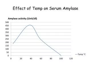
Susithra Selvam _____________________________________________________________________________ Der Pharma Chemica, 2015, 7 (8):59-63 tryptophan (Trp) moiety, which is fluorescent in nature, fluorescence based spectroscopic analysis on the association of BSA with NaC media is carried out. MATERIALS AND METHODS Bovine serum albumin was purchased from Sigma chemicals, USA and sodium cholate was purchased from Sisco Research Laboratories Private Limited (India). Both chemicals were used without any further purification. A 50 mM sodium phosphate buffer of pH 7.4 was used for all experiments. A Horiba Jobin-Yvon Fluoromax-4 spectrofluorometer was used to record the fluorescence spectra. Excitation and emission spectra were measured with 2 nm band width. The steady state fluorescence anisotropy is defined as [19], I -GI r =I +2GI Where, IVV and IVH are the fluorescence intensities and the subscript indicates the vertical (V) and horizontal (H) orientations of the excitation and emission polarizer. G is the instrumental correction factor: I G= I Preparation of BSA - NaC solutions The stock solutions of NaC were prepared in 50 mM phosphate buffer pH 7.4 at concentration higher, than the critical micellar concentration (cmc) (NaC = 6-16 mM). A range of different concentrations of bile salt solutions are prepared by diluting these stock solutions using buffer solution. BSA concentration was maintained at 10 µM for all the experiments. The range of concentration of bile salt varied is NaC = 4.8 – 43.2 mM. The solutions were incubated for 2 hrs to achieve equilibrium. RESULTS AND DISCUSSION Fluorescence emission Spectra of BSA in presence of NaC The association of BSA with the bile salts, namely NaC was studied using the absorption and fluorescence property of tryptophan (Trp) moiety, with an excitation at 290 nm and emission at 350 nm. With the initial addition of NaC, the Trp fluorescence is quenched upto ~ 50 % (Figure 1)20,21 and after that there is gradual, but small increase in the fluorescence intensity of BSA is observed with increasing concentration of NaC. Figure 1. Emission spectra of BSA (λex 290 nm) with NaC. pH = 7.4, T = 25 °C The quenching of Trp fluorescence clearly indicates that the association of bile salts with the protein is at a close proximity to the Trp moiety. There is a slight blue shift of Trp fluorescence with increasing concentration of bile VV VH ss VV VH HV HH 60 www.scholarsresearchlibrary.com
Susithra Selvam _____________________________________________________________________________ Der Pharma Chemica, 2015, 7 (8):59-63 salts. This indicates that the environment of Trp moiety is becoming more hydrophobic with the addition of bile salts. The data also reveals that the Trp fluorescence quenched with initial addition of bile salt and with increasing bile salt concentration, Trp fluorescence intensity increases gradually (Figure 2A). The interpretation of the data reveals that the change in Trp fluorescence after the initial addition of BSA is small and it can be proposed that NaC molecules favor the stabilization of serum albumins. Figure 2. Plot of variation of Trp fluorescence intensity (λex 290 nm; λem 350 nm) with increasing concentration of NaC. (A) [NaC] = 0 – 43.2 mM and (B) [NaC] = 4.82 – 43.2 mM. pH = 7.4, T = 25 °C Even though there is increase in Trp fluorescence intensity, the change is minimal, but not negligible. The fluorescence emission property of Trp moiety lies closer to the NaC aggregates and this enhancement of Trp emission follows the aggregation pattern of bile salts; viz primary dimers are formed initially and then the secondary larger aggregates, with the cmc range at 6 – 16 mM for NaC. The study shows that Trp fluorescence is not only effective in understanding the association of BSA with NaC, but also capable of sensing the micellization of bile salts. The hydrophobic nature of BSA – NaC is clearly indicated by the Trp emission intensity enhancement with increasing concentration of NaC. Steady state fluorescence anisotropy of BSA-NaC system Figure 3 gives the steady state fluorescence anisotropy (rss) of BSA in presence of increasing concentration of NaC. The fluorescence anisotropy of Trp shows a slight increase from 0.09 to 0.11 with increasing concentration of NaC. 61 www.scholarsresearchlibrary.com
Susithra Selvam _____________________________________________________________________________ Der Pharma Chemica, 2015, 7 (8):59-63 The slight increase in fluorescence anisotropy is again an indicative of stabilization of BSA in their native state by NaC media. Figure 3. Plot of variation of fluorescence anisotropy of Trp (λex 290 nm; λem 350 nm) with increasing concentration of NaC. [NaC] = 0 – 43.2 mM. pH = 7.4, T = 25 °C Also figure 3 gives a clear analysis of the Trp fluorescence following the aggregation pattern of NaC molecules. Further the rss values suggest that the association of BSA with bile salts is hydrophobic in nature. Association of BSA with NaC aggregates The Trp fluorescence emission and fluorescence anisotropy studies of the association of BSA with NaC show that the initial addition of NaC molecule quenches the Trp fluorescence of about 50 %. A similar effect was observed in literature, where the quenching was observed in the lower concentrations of NaC [20,21]. This indicates the NaC being incorporated into protein macromolecule, with a close proximity to the Trp moiety. BSA has two Trp moieties viz. Trp 212, found in the hydrophobic core of the serum albumin with the sub-domain IIA and Trp 134 in the sub- domain IA, which is exposed to solvent. Further addition of NaC increases the Trp fluorescence to a small extent. Even though the increase is smaller, it follows the aggregation pattern of NaC media through its cmc range, viz. 6 to 16 mM (figure 2). The steady state fluorescence anisotropy study of Trp fluorescence is in collaboration with the fluorescence emission of BSA in NaC media. Both the studies clearly indicate that association of BSA with NaC media is hydrophobic in nature and also that bile salt media can be used as stabilizing agent for BSA to preserve them in their native state. CONCLUSION The association of serum albumin, BSA with the bile salt, NaC was studied through the Trp fluorescence emission and fluorescence anisotropy. A quenching of Trp fluorescence is observed with the initial addition of NaC to BSA and further small increase in Trp fluorescence is seen with increasing concentration of NaC. The data is indicative of NaC being incorporated within the BSA at a close proximity to the Trp moiety and that NaC media being capable of stabilizing protein in their native state. A similar observation is made from the fluorescence anisotropy studies. Thus it can be concluded that the nature of interaction between BSA and NaC moieties is hydrophobic and NaC media can be used as a pharmaceutical excipient to stabilize the BSA in their native state. Acknowledgements The author, Dr. Susithra Selvam is thankful to DST – SERB, India (SERB/F/2979/2013-14) for the financial support. The author also thanks Prof. Ashok Kumar Mishra, Department of Chemistry, Indian Institute Technology of Madras for unrestricted usage of spectrofluorometer. 0.112 0.110 0.108 0.106 Fl. Anisotropy 0.104 0.102 0.100 0.098 0.096 0.094 0.092 0 10 20 30 40 50 [NaC] (mM) 62 www.scholarsresearchlibrary.com
Susithra Selvam _____________________________________________________________________________ Der Pharma Chemica, 2015, 7 (8):59-63 REFERENCES [1]Kumar, V.; Sharma, V.K.; Kalonia D.S. Int. J. Pharm.2005, 294, 193-199. [2]Ruiz, C.C.; Hierrezuelo, J.M.; Peula-Garcia, J.M.; Aguiar, J. The open macromolecules journal, 2008, 2, 6 – 18. [3]Recalde Ruiz, D.L.; Carvhalo Torres, A.L.; Andres Garcia, E.; Diaz Garcia M.E. Analyst1998, 123, 2257 – 2261. [4]Deo, N.; Jockusch, S.; Turro, N.J.; Somasundaran P. Langmuir, 2003, 19, 5083 – 5088. [5]Togashi, D.M.; Ryder, A.G. J. Fluoresc.2006, 16, 153 – 160. [6]Maurer, N.; Fenske, D.B.; Cullis, P.R. Expert Opin. Biol. Ther.2001, 1, 1 – 25. [7]Farokhzad, O.C.; Langer, R. ACS Nano. 2009, 3, 16 – 20. [8]Abu Lila, A.S.; Ishida, T.; Kiwada, H. Expert Opin. Drug Deliv.2009, 6, 1297 – 1309. [9]Maldonado-Valderrama, J.; Wilde, P.; Macierzanka, A.; Mackie, A. Adv. Colloid Interface Sci. 2011, 165, 36 – 46. [10]O’Connor, C.; Wallace R.G. Adv. Colloids Interface Sci.1985, 22, 1 – 111. [11]Mukhopadhyay, S.; Maitra, U. Curr. Sci.2004, 87, 1666 – 1683. [12]Small, D.M; Penkett, S.A.; Chapman, D. Biochim. Biophys. Acta1969, 176, 178 - 189. [13]Reis, S.; Moutinho, C.G.; Matosa, C.; de Castro, B.; Gameiroc, P.; Lima, J.L.F.C. Anal. Biochem.2004, 334, 117 – 126. [14]Susithra, S.; Maneesha E.A.; Mishra A.K. J. Pharm. Sci.2009, 98, 4153 - 4160. [15]Wang, W. Int. J. Pharm. 2005, 289, 1 – 30. [16]Crommelin, D.J.A.; Jiskoot, W. Eur. J. Hosp. Pharm. Prac.2006, 12, 14 – 26. [17]Tana, M.L.; Choonga, P.L.M.; Dassa, C.R. Peptides2010, 31, 184 – 193. [18]Dai, C.; Wanga, B.; Zhao, H. Colloids Surf., B.2005, 41, 117 – 120. [19]Lakowicz, J.R. Principles of fluorescence spectroscopy. Springer 3rd ed, New York, 2006. [20]De, S.; Das, S.; Girigoswami, A. Colloid. Surface. B2007, 54, 74 – 81. [21]De, S.; Girigoswami, A.; Das, S. J. Colloid Interf. Sci.2005, 285, 562 – 573. 63 www.scholarsresearchlibrary.com