Download
1 / 14
140 likes | 162 Vues
Pseudo first order reaction kinetics and binding studies of trans-[Co(en)2(CH3CH2CH2)H2O]2 complex with<br>imidazole ,substituted imidazoles, glycine and ethyl glycine ester has been investigated using spectrophotometric<br>technique. Equilibrium constants were determined as a function of pH at 25°c. Binding and kinetic studies were<br>correlated based on basicity, steric hindrance. From the equilibrium data, it is found that the entering nucleophile is<br>participating in the transition state, thereby SN1<br> mechanism is proposed. The effect of the incoming ligands on the<br>complex is studied by molecular mechanics. The interaction of trans-[Co(en)2(CH3CH2CH2)H2O]2 with CT DNA<br>has been studied spectrophotometrically. <br>

E N D
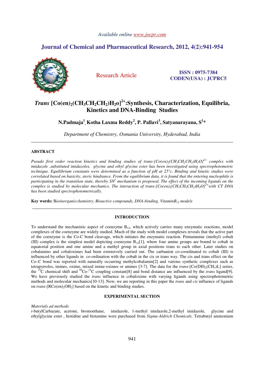
Available online www.jocpr.com Journal of Chemical and Pharmaceutical Research, 2012, 4(2):941-954 ISSN : 0975-7384 CODEN(USA) : JCPRC5 Research Article Trans [Co(en)2(CH3CH2CH2)H2o]2+:Synthesis, Characterization, Equilibria, Kinetics and DNA-Binding Studies N.Padmaja1, Kotha Laxma Reddy2, P. Pallavi3, Satyanarayana, S1* Department of Chemistry, Osmania University, Hyderabad, India ______________________________________________________________________________ ABSTRACT Pseudo first order reaction kinetics and binding studies of trans-[Co(en)2(CH3CH2CH2)H2O]2+ complex with imidazole ,substituted imidazoles, glycine and ethyl glycine ester has been investigated using spectrophotometric technique. Equilibrium constants were determined as a function of pH at 25°c. Binding and kinetic studies were correlated based on basicity, steric hindrance. From the equilibrium data, it is found that the entering nucleophile is participating in the transition state, thereby SN1 mechanism is proposed. The effect of the incoming ligands on the complex is studied by molecular mechanics. The interaction of trans-[Co(en)2(CH3CH2CH2)H2O]2+with CT DNA has been studied spectrophotometrically. Key words: Bioinorganicchemistry, Bioactive compounds, DNA-binding, VitaminB12 models _____________________________________________________________________________ INTRODUCTION To understand the mechanistic aspect of coenzyme B12, which actively carries many enzymatic reactions, model complexes of the coenzyme are widely studied. Much of the study with model complexes reveals that the active part of the coenzyme is the Co-C bond cleavage, which initiates the enzymatic reaction. Pentammine (methyl) cobalt (III) complex is the simplest model depicting coenzyme B12[1], where four amine groups are bound to cobalt in equatorial position and one amine and a methyl group in axial positions trans to each other. Later studies on cobalamins and cobaloximes had been extensively carried out. The carbanion co-coordinated to cobalt (III) is influenced by other ligands in co-ordination with the cobalt in the cis or trans way. The cis and trans effect on the Co–C bond was reported with naturally occurring methylcobalamin[2] and various synthetic complexes such as tetrapyrroles, imines, oxime, mixed imine-oximes or amines [3-7]. The data for the trans-[Co(DH)2(CH3)L] series, the 13C chemical shift and 59Co-13C coupling constant[8] and bond distance are influenced by the trans ligand[9]. We have previously studied the trans influence in cobaloxime with varying ligands using spectrophotometric methods and molecular mechanics[10-13]. Now, we are reporting in this paper the trans and cis influence of ligands on trans-[RCo(en)2OH2] based on the kinetic and binding studies. EXPERIMENTAL SECTION Materials ad methods t-butylCarbazate, acetone, bromoethane, imidazole, 1-methyl imidazole,2-methyl imidazole, glycine and ethylglycine ester , histidine and histamine were purchased from Sigma-Aldrich Chemicals. Tetrabutyl ammonium 941
Satyanarayana, Set al ______________________________________________________________________________ J. Chem. Pharm. Res., 2012, 4(2):941-954 hydrogen sulfate (TBAHS), potassium hydroxide, magnesium sulfate, conc. HCl, cobalt nitrate, ammonia solution and methanol were obtained from Merck. Synthesis of complex Trans-[Co(en)2(CH3CH2CH2)H2O]2+has been prepared according to the previously available literature[1][7b][ found C, 21.035; H, 6.532; N, 14.312. Calc. for C7N4 H25 O1 Co: C, 21.010; H, 6.297; N, 14.0023 %] UV-Vis peaks at;245,306,437 as given in Fig 1;IR: 1458 (C=C), 1578 (C=N), 80 (Co–N (en)), 578 (Co–N (L))as given in Fig 2. 1H –NMR as given in Fig 3, 13C-NMRas given in Fig.4,LC-MS: m/z= 398(APCI-NEG1) in Fig.5, DTA-TG- MS in Fig 6 andmolecularmodelingstructures in Fig 9,10,11. The Propylhydrazine was prepared[14]and as it is used as an alkylating agent. The Scheme 1 shows the synthesis. Physical measurements Elemental analyses (C,H and N) was performed on Flash EA 1112 series Thermofinnigon. The pH of the solution was measured by a DigisunpH meter DI-707. For the calibration of the pH meter, standard buffers of pH 4.0,7.0 and 9.2 were used. Mass spectra was recorded on LC-MS- 2010A Schimadzu with Column- C-18,Detector-UV(254) and MS probe of ESI. Infrared spectra was recorded on a Perkin –Elmer 1600 series-FTIR Spectrometer as KBr discs. NMR spectra on a Bruker ARX-300 NMR Spectrometer with D2O as a solvent. TG8110thermal analyzer was used to record simultaneous TG,DTA,MS curves in the temperature range of 20-8000c using platinum crucibles. UV-VIS spectra were recorded on a Elico BL 198model spectrophotometer with temperature control, models for molecules are drawn with the help of Hyperchem software and energies are calculated using the same software. Kinetics and binding studies were performed on a Elico SL 171 model single beam spectrophotometer. Spectroscopic characterization Complex was characterized by UV-Vis peaks at; 245,306,437; IR: 1458 (C=C), 1578 (C=N), 80 Co–N (en)), 578 (Co–N (L)). 1H –NMR peaks at2.825(t)(CH3), 3.12(m)(CH2),3.14(t)(CH2) and 2.63(t) (CH2en) ppm, 13C-NMR peaks at40.48(CH2), 39.31(CH3),44.5(CH2en) ppm, and LC-MS: (APCI-NEG1) studies with column- C- 18,Detector-UV(254) and MS probe of ESI shows that the calculated m/z value was matching with the recorded m/z value (m/z 398). Thecharacterization of the structure and energies of molecular complexes are essential for understanding many biological functions. To be able to predict the strength of non covalent bonding between molecular and 3D structures of the corresponding complexes has thus been along standing goal in computational chemistry significant progress has been made in computer aided ligand design during the past decade and methodologies based on force field calculations such as Molecular Mechanics, Molecular Dynamics and Monte Carlo simulations have been important for many of these developments [28-31]. Molecular mechanistic studies were performed on complex. The optimized structure of the complex is given in Fig (9),all bond lengths and bond angles are given in table (4 & 5) Thermal analysis The thermal analysis curves (TG, DTA, MS) of the studied complex was shown in Fig (6). The Thermogravimetry (TG) and Differential thermal analysis curves showed a three step decomposition pattern. In the first step the on set temp (Ton ) at 231.60c and end set temp(Ten ) at 259.90c has been attributed to the loss of water molecule with a mass change of -17.65%. In the second step theon set temp (Ton ) at 350.90cand end set temp(Ten )at 387.80chas been attributed to the mass change of -12.28%. In the third step the on set temp (Ton ) at 672.10cand end set temp (Ten)at 743.70chas been attributed to the mass change of -21.58%. In the DTA curve, there are three peaks. The first endothermic peak appears at 249.60c, the second endothermic peak appears at 369.40c and the third endothermic peak appears at 707.90c. At the end of decomposition cobalt oxide is present. Binding and Kinetic Studies The binding and kinetic studies were carried out using an Elico single beam spectrophotometer (SL-171 model). Spectra were recorded on ElicoBL198 Model. The concentrations were fixed at 480nm. The sample compartment temperature was maintained at25° ?1°c. Determination of Equilibrium Constant The apparent binding constants (Kapp) were [Co(en)2(CH3CH2CH2)H2O]2+complex with different ligands. By taking fixed concentration of complex and by varying the ligand concentration the absorbance was recorded. Solutions containing an appropriate buffer (0.2M) to determined for the axial ligation of trans- 942
Satyanarayana, Set al ______________________________________________________________________________ J. Chem. Pharm. Res., 2012, 4(2):941-954 maintain pH, KCl to maintain ionic strength (1.0M), varying concentrations of ligand are taken in a 3 ml cuvette and allowed to equilibrate in a thermostat cell holder at 25?0.1°c for 15 min prior to the addition of trans- [Co(en)2(CH3CH2CH2)H2O]2+. Absorbance’s were recorded and the apparent equilibrium constants were calculated from the plot of ∆A/[L]fvs.∆A. Thus, for each ligand Kapp was calculated using Eq. 1. ∆A = ∆Amax [L]f / (1/Kapp + [L]f) ---------------- (1) The least square fit of the above equation after rearrangement is given by Eq. 2: ∆A = ∆Amax – {1 / Kapp (∆A / [L]f)} ----------------- (2) Where ∆A = difference in absorbance between solutions containing only complex with and without ligand. ∆Amax = maximum difference in absorbance recorded at high ligand concentration. [L]f = the unbound ligand concentration and is calculated from Eq. 3 [L]f= [L]T – (CT∆A / ∆Amax) ------------------- (3) [L]T = the total volume of ligand added CT = the total concentration of trans-[Co(en)2(CH3CH2CH2)H2O]2+ The pH independent equilibrium constants are then calculated from Eq. 4: Keq = Kapp / aL --------------- - (4) Where aL (fraction of ligand as free base) was calculated from Eq. 5: aL = Ka / (Ka + [H+]) ------------------- (5) Kinetic Studies Wehave investigated the ligand substitution reactions of trans-[Co(en)2(CH3CH2CH2)H2O]2+with imidazole, substituted imidazoles at 25°c. The reaction rates were determined by maintaining pseudo-first order conditions by taking 10-fold excess of ligand concentration with respect to the complex concentration. The kinetics was studied by varying the concentration of the ligand at 486nm and using appropriate buffer at pH below the pKa of the ligand. The absorbance was monitored at λmax 480 nm. The first order rate constants (kobs) are obtained by least square fits of the data to Eq. 6 below. lnAt – ln A∞ = kobst ---------------------- (6) Where Atis the absorbance at time‘t’ and A∞ is the final absorbance. RESULTS AND DISCUSSION The ligand substitution reactions of trans-[Co(en)2(CH3CH2CH2)H2O]2+with imidazole, substituted imidazoles,histidine, histamine, glycine and ethyl glycine esterare given . The UV-Vis scan of trans- [Co(en)2(CH3CH2CH2)H2O]2+is given in Fig. 1. Depending on the pKa values of the ligands, the binding studies are made in the pH range above and below the pKa values. The Kapp values were determined as a function of pH by spectrophotometry. The dependence of logKapp for ligation of trans-[Co(en)2(CH3CH2CH2)H2O]2+with different ligands upon pH is given in Fig. 7 and the data given in Table 1. Up to pKa of the ligand, the log Kapp increases with pH but above pKa the log Kapp is independent of pH. It is observed that the Kapp value below the pKa value is very low due to the protonation of the ligand and as the pH increases, ligand gets deprotonated and binds strongly to Co 943
Satyanarayana, Set al ______________________________________________________________________________ J. Chem. Pharm. Res., 2012, 4(2):941-954 (III) and Kapp increases. The Keq values for the binding of Imidazoles to [Co(en)2(CH3CH2CH2)H2O]2+follow the order: 1-MeIMD > IMD > 2-MeIMD Where as the binding of aminoacids follow the order Glycine>Glycine ethyl ester The stability order can be explained by considering the HSAB principle, basicity of ligands and their ability of π- bonding and σ-donation. Considering imidazole series for 1-MeIMD and IMD the formation constants are high for higher pKa valuesi.e. they follow the basicity order. Though 2-Me-Imidazole is more basic than Imidazole, the Keq is smaller. This can be attributed to the steric hindrance due to the preference of methyl group at C2position. Among glycine and glycine ester, both are σ-donors but glycine has more binding constant as it is more basic than ethyl glycine ester. Though amino acids are more basic than Imidazoles they form less stable complexes. The stability order of Imidazoles is attributed to the ability of imidazoles to bind with Co(III) through dΠ-pΠ back bonding. Glycine and ethyl glycine ester are only σ donors,cannot accept electrons in a similar way. The rate of ligand substitution is pH dependent. The rate of the reaction increases drastically near the pKa of the ligand. The slope of the plot of kobsvs. concentration of the ligands is given in Fig (8) and the data for the plot is given in Table 2. The comparison of second order rate constantkon’ at a given pH is given in table 3. The slopes of the least square fit of the Eq. 7 gives the second order rate constant. kobs = kon’[L]T + koff ---------------------------- (7) [L]T =Total ligand concentration The pH independent second order rate constants, kon are obtained by using the Eq. 8. kon = kon’ / aL ------------------------------- (8) The second order rate constants increase as the nucleophilicity of the ligand increases. This is in accordance with the order of Keq values. The kinetics of substitution of the axial base in alkylcobaloximes and related cobalt complexes has been studied under a variety of conditions[15,16]. The studies on cobalt complexes and adenosylcobaloxime provide evidences for the mechanism of substitution to be dissociative[16,17] (Id or D). In view of the evidence presented above, for the existence of pentacoordinate alkylcobaloximes and the ligation kinetic studies of others, both on alkyl cobalt complexes and on cobaloxime complexes [18,19] with other equatorial ligand system[20], an SN1 mechanism may be suggested. Molecular Mechanistic Studies The structural investigation of coordination and organometallic chemistry has been advanced using molecular mechanics[21-24]. Using MM2 parameterization, the optimized structure was deduced using Hyperchem software inFig.9 shows ball and stick representation of complex.The optimized structure of Complex with imidazole has been given in Fig 10, the Complex with Glycine has been given in Fig 11. Bond lengths and bond angles are given in tables 4 & 5. DNA Binding. Absorption Spectral Studies The application of electronic absorption spectroscopy in DNA-binding studies is one of the most useful techniques[25, 26] metal complex binding with DNA through groove mode usually results in hypochromism and bathochromism, due to the groove mode involving a strong stacking interaction between an aromatic chromophore and the base pairs of DNA. The extent of the hypochromism commonly parallels the groove binding strength. The absorption spectra of the complex in absence and presence of calf thymus DNA using Tris buffer are illustrated in Fig. 12 . In the UV region, the intense absorption bands observed in Co(III) complexes are attributed to intraligand dΠ–pΠtransition of the coordinated groups. Addition of increasing amounts of CT DNA results in hypochromism and moderate bathochromic shift in the UV spectra of the complex -[Co(en)2(CH3CH2CH2)H2O]2+. These spectral data 944
Satyanarayana, Set al ______________________________________________________________________________ J. Chem. Pharm. Res., 2012, 4(2):941-954 may suggest a mode of binding that involves a stacking interaction between the complex and the base pairs of DNA. In order to quantitatively compare the binding strength of the two complexes, the intrinsic binding constants K of the complexes with CT DNA were determined according to the following equation [27] through a plot of [DNA] / (åb- åf) vs. [DNA] (Eq. 9). [DNA] / (åa – åf) = [DNA] / (åb – åf) + 1/(K (åb – åf)) (9) Where [DNA] is the concentration of DNA in base pairs, the apparent absorption coefficient åa , åf and åb correspond to Aobs / [Co], the extinction coefficient for the cobalt complex in the free and fully bound form, respectively. In plots DNA] / (åb-åf) vs. [DNA]. K is given by the ratio of slope to intercept. DNA binding constantK was about 1.6 x 10⁴ Mrespectively from the decay of the absorbance. The binding constants indicate that the complex binds more strongly. Fluorescence Studies The complexes can emit luminescence in Tris buffer (pH 7.0) with the emission maxima at 619nm. Binding of complexto DNA was found to increase the fluorescence intensity. The emission spectra of complex in the absence and presence of CT DNA are shown in Fig 13. The plots of the emission quenching intensity versus the ratio of [DNA]/[Co] are also inserted in Fig 14. Upon addition of CT DNA, the emission intensity increases steadily. The increase in emission intensity of complex is that these results were strengthened by viscosity studies. Table1: Formation constants(Kapp) for the axial ligation of the aqua bis (ethylene diamine) propyl cobalt(III) complex by different ligands at 250 C for different pH Values pH 1-MeImd Imd 2-MeImd 5.0 – 5.5 – 6.0 1.456 1.32 0.563 6.5 1.916 1.77 1.052 7.0 2.312 2.144 1.521 7.5 2.59 2.39 1.93 8.0 2.735 2.512 2.225 8.5 2.793 2.56 2.38 9.0 2.813 2.57 2.45 10.0 2.821 2.58 2.48 11.0 2.823 2.58 2.48 Keq 664.31 381.54 304.51 Table 2 : Dependence of the rate constants (kobs) for the axial ligation of aqua bis(ethylene diamine) propyl cobalt( III ) on the concentration of the ligand at 250 C M/L 1-MeImd Imd 2-MeImd Gly – – 0.203 0.697 1.153 1.635 2.26 2.424 280 Gly-OEt 0.556 1.044 1.484 1.832 2.05 1.145 2.18 2.2 158 Hist 1.27 2.1 2.34 2.4 2.5 2.52 2.52 2.53 2.53 2.53 336.88 Histmn 1.09 1.57 1.96 2.26 2.42 2.5 2.5 2.5 2.5 2.5 2.5 328.68 Gly 2×10⁻⁵ (1:10) 3×10⁻⁵ (1:20) 7×10⁻⁵ (1:30) 9×10⁻⁵ (1:50) 9x10⁻⁵ 11.0 0.948 9.5×10⁻⁵ Gly-Oet 2×10⁻⁵ (1:10) 2×10⁻⁵ (1:20) 2×10⁻⁵ (1:30) 5×10⁻⁵ (1:50) 8×10⁻⁵ 8.0 0.707 1.1×10⁻⁴ Hist 2×10⁻⁵ (1:10) 2×10⁻⁵ (1:20) 2×10⁻⁵ (1:30) 4×10⁻⁵ (1:50) 2.82x10⁻⁴ 9.0 0.856 3.3x10⁻⁴ Histmn 2×10⁻⁵ (1:10) 2×10⁻⁵ (1:20) 2×10⁻⁵ (1:30) 4×10⁻⁵ (1:50) 2.64x10⁻⁴ 9.0 0.856 3.1x10⁻⁴ kobs (S-1) 1:50 2×10⁻⁵ 1×10⁻⁵ 2×10⁻⁵ 1:100 2×10⁻⁵ 2×10⁻⁵ 2×10⁻⁵ 1:150 2×10⁻⁵ 3×10⁻⁵ 6×10⁻⁵ 1:200 5×10⁻⁵ 3×10⁻⁵ 8×10⁻⁵ 3.62x10⁻⁴ 7.5 0.586 6.17x10⁻⁴ 3x10⁻⁴ 7.5 0.645 4.65x10⁻⁴ 2.3x10⁻⁴ 7.5 0.28 8.2×10⁻⁴ kon’ pH α kon (dm3mol-1sec-1) Table 3 : Comparison of second order rate constants kon for the formation of [CH3CH2CH2Co(en)2OH2] 2+at 250C obtained from trans aquo and amino cobalt (III) complex. Complexes 1-MeImd Imd [CH3CH2CH2Co(en)2OH2] 2+ 6.17x10⁻⁴ 4.65x10⁻⁴ 2-MeImd 8.2×10⁻⁴ Gly Gly-Oet 1.1×10⁻⁴ Hist 3.3x10⁻⁴ Histmn 3.1x10⁻⁴ 9.5×10⁻⁵ 945
Satyanarayana, Set al ______________________________________________________________________________ J. Chem. Pharm. Res., 2012, 4(2):941-954 Table- 4: Bond lengths of [CH3CH2CH2Co(en)2L] 2+ 1-Me- Imidazole 2.17804 2.20704 2.08436 1.72366 2.1604 2.1604 1.4939 1.46364 1.56896 1.72434 1.8443 1.3727 1.0804 1.58446 2-Me- Imidazole 2.2869 2.2047 1.92763 1.92579 2.26403 2.3734 1.5492 1.54518 1.5022 1.53297 1.49789 1.58613 1.67774 1.61282 Ethyl glycine ester 1.9112 1.88635 1.8838 1.89938 1.9667 1.8962 1.46720 1.4681 1.47107 1.4747 1.49767 1.50315 1.5313 1.5319 Complex Imidazole Glycine Histidine Histamine [CH3CH2CH2Co(en)2OH2] 2+ Co1- N2 Co1- N3 Co1- N4 Co1- N5 Co1- C10 Co1- N13 C6- N2 C7- N3 C8- N4 C9- N5 C6- C7 C8- C9 C10 -C11 C11 -C12 2.2421 2.05996 2.32152 1.8615 2.11777 2.33593 1.5252 1.5438 1.62573 1.49938 1.50174 1.55662 1.61806 1.5863 1.9005 1.9112 1.9092 1.9041 1.9642 1.8495 1.4792 1.4688 1.4717 1.4712 1.5263 1.532 1.5532 1.5424 2.19146 2.0337 2.0676 1.8622 2.0968 1.9053 1.52573 1.5403 1.5745 1.66001 1.8587 1.5544 1.5902 1.5821 1.88312 1.8859 1.9009 1.9012 1.95606 2.3756 1.4678 1.4678 1.4691 1.4638 1.5054 1.4999 1.5368 1.53035 Table- 5: Bond angles of [CH3CH2CH2Co(en)2L] 2+ Complex Imidazole 72.5564 35.093 118.554 144.798 125.121 124.499 106.009 65.1412 103.639 114.184 157.949 1-Me- Imidazole 51.9972 85.8293 124.958 122.872 141.69 144.923 98.1917 108.149 102.803 98.5872 159.784 2-Me-Imidazole 166.254 37.703 126.239 165.364 123.24 129.043 106.57 68.3373 106.57 113.964 166.254 Glycine 85.2259 85.4454 110.93 112.255 113.607 110.844 109.885 104.673 110.526 107.153 129.991 Ethyl glycine ester 50.0819 66.6631 110.853 111.487 112.014 112.014 102.085 104.78 101.061 115.231 159.87 Histidine 70.822 81.7452 127.503 125.273 128.453 126.621 98.978 106.502 101.377 112.728 150.721 Histamine 72.72 84.992 123.202 109.11 111.634 110.311 106.693 105.50 111.908 104.33 115.908 [CH3CH2CH2Co(en)2OH2] 2+ 4-1-8 5-1-9 8-4-1 1-5-9 6-2-1 7-3-1 4-8-9 5-9-8 2-6-7 3-7-6 10-1-13 Fig 1:U.V.Visible scan of [CH3CH2CH2Co(en)2OH2] 2+ This observation is further supported by the emission quenching experiments using [Fe(CN)6]4- as a quencher. The ion [Fe(CN)6]4- has been shown to be able to distinguish differentially bound cobalt (III) species and positively charged free complex ions. The complex binding to DNA can be protected from the quencher, because highly negatively charged [Fe(CN)6]4- would be repelled by the negative DNA phosphate backbone, hindering quenching of the emission of the bound complex. The method essentially consists of titrating a given amount of DNA-metal complexes with increasing concentrations of [Fe(CN)6]4- and measuring the change in fluorescence intensity. The 946
Satyanarayana, Set al ______________________________________________________________________________ J. Chem. Pharm. Res., 2012, 4(2):941-954 ferro-cyanide quenching curves of the complex in the presence and absence of CT DNA are shown in Fig (14). The absorption and fluorescence spectroscopy studies determine the binding of complexes with DNA. Fig2: IR Spectrum of [CH3CH2CH2Co(en)2OH2] 2+ Fig3: 1H NMR Spectrum of [CH3CH2CH2Co(en)2OH2] 2+ 947
Satyanarayana, Set al ______________________________________________________________________________ J. Chem. Pharm. Res., 2012, 4(2):941-954 Fig 4 : 13C [1H] NMR Spectrum of [CH3CH2CH2Co(en)2OH2] 2+ Fig 5 : LC- MS Spectrum of [CH3CH2CH2Co(en)2OH2] 2+ 948
Satyanarayana, Set al ______________________________________________________________________________ ______________________________________________________________________________ ______________________________________________________________________________ J. Chem. Pharm. Res., 2012, 4(2): J. Chem. Pharm. Res., 2012, 4(2):941-954 Fig 6 : Thermogram of 6 : Thermogram of[CH3CH2CH2Co(en)2OH2] 2+ Fig 7: Dependence of formation constants ( logKapp) on the pH for the axial ligation of Fig 7: Dependence of formation constants ( logK Co(en)2H2O]2+ by different ligands (L) at 25 by different ligands (L) at 250C in aqueous solution, ionic strength 1.0 M KCl. ) on the pH for the axial ligation of trans-[CH3 CH2CH2 C in aqueous solution, ionic strength 1.0 M KCl. 949
Satyanarayana, Set al ______________________________________________________________________________ ______________________________________________________________________________ ______________________________________________________________________________ J. Chem. Pharm. Res., 2012, 4(2): J. Chem. Pharm. Res., 2012, 4(2):941-954 [CH3 CH2CH2 Co(en)2 H2O] Fig 8: Dependence of the Rate Constants (k 2+on the Concentration of different ligands (L) at 25 8: Dependence of the Rate Constants (kobs) for the Axial Ligation of trans- [CH different ligands (L) at 250C in aqueous solution, ionic strength 1.0 M KCl. C in aqueous solution, ionic strength 1.0 M KCl. Fig. 9: Aqua bis (ethane Fig. 9: Aqua bis (ethane -1,2-diamine) propyl Cobalt (III) complex propyl Cobalt (III) complex 950
Satyanarayana, Set al ______________________________________________________________________________ J. Chem. Pharm. Res., 2012, 4(2):941-954 Fig 10: Imidazole (ethane – 1,2-diamine) propyl Coblat(III) Complex Fig 11: Glycine (ethane – 1,2-diamine) propyl Coblat(III) Complex 951
Satyanarayana, Set al ______________________________________________________________________________ J. Chem. Pharm. Res., 2012, 4(2):941-954 Fig - 12 U.V.Visible scan of DNA binding of [CH3 CH2CH2 Co(en)2H2O] 2+ 130 y = 0.0332x - 2E-08 1.40E-06 1.20E-06 1.00E-06 8.00E-07 Io /I 6.00E-07 100 4.00E-07 2.00E-07 0.00E+00 0.00E+0 0 1.00E-05 2.00E-05 3.00E-05 4.00E-05 [Fe(CN)6]4- Int. 50 0 540 550 560 570 577 Wavelength[nm] Fig -13:Fluorescente emisión spectra of complex[CH3 CH2CH2 Co(en)2H2O]2+ in aqueous buffer. Tris 5mM, NaCl 50mM, pH 7.0) in the presence of CT DNA, [Co] = 20µM, [DNA] / [Co] 0,5,10,15,20 (The arrow shows the intensity changes upon increasing concentration. Inset: Plots of relative integrated emission intensity vs [DNA] / [Co]. 952
Satyanarayana, Set al ______________________________________________________________________________ J. Chem. Pharm. Res., 2012, 4(2):941-954 9 8 7 6 5 4 3 2 1 0 0 0.02 0.04 0.06 0.08 [ Fe( CN) 6] 4- Fig 14 :Emission quenching curves of [CH3 CH2CH2 Co(en)2H2O] 2+in the absence ofof DNA (upper) and presence of DNA (lower) CONCLUSION The ligand substitution reactions were studied by different ligands on trans-aquo bis(ethylenediamine)Propyl cobalt(III) andfollowthe basicity order 1-MeIMD>IMD>2-MeIMD>glycine>glycine ethyl ester. The binding and kinetic constants varied with the incoming nucleophile suggesting that the nucleophile is taking part in the transition state. Thus, Id mechanism is suggested. The DNA binding studies suggests that the complex binds with CTDNA. Acknowledgement We gratefully acknowledge the UGC, New Delhi for financial support in the form of major research project. REFERENCES [1]P. Kofod, Inorg.Chem. 1995,34, 2768. [2]Eds: B. Kräutler, D. Arigoni, B. T. Golding, ‘Vitamin B12 and B12-Proteins’, Wiley-VCH, Weinheim, Germany, 1998. [3]D. Dolphin, A. W. Johnson, Chem. Commun. (London) 1965, 494. [4]K. Farmery, D. H. Busch, Inorg Chem, 1972, 11, 2901. [5]G. N. Schrauzer, Angew. Chem., Int. Ed.1976, 15,417; [6]B. D. Gupta, V. Vijaikanth, V. Singh, Organometallics, 2004, 23, 2069; [7]V. Vijaikanth, B. D. Gupta, D. Mandal, S. Shekhar, Organometallics 2005, 24,4454. [8]G. Costa, G. Mestroni, E. de Savorgnami, Inorg. Chim. Acta1969, 3, 323. a) T. S. Roche, J. F. Endicott, J. Am. Chem. Soc.1972, 94, 8622.; b) P. Kofod, P. Harris, S. Larsen, Inorg.Chem. 1997, 36, 2258. [9]F. Asaro, L. Liguori, G. Pellizer, Phys. Chem. Chem. Phys.1999, 1, 4981. [10]L. Randaccio, N. Bresciani-Pahor, E. Zangrando, L. G. Marzilli, Chem. Soc. Rev.1989, 18, 225. [11]D. R. Sudharshan, B. R. Krishna, P. Pallavi, N. Navaneetha, S. Satyanarayana, Indian. J. of Chem.2005, 44A, 678. [12]N. Navaneetha, S. Satyanarayana, Indian J. of Chem. 2005, 44A, 1191. [13]J. V. Madhuri, V. Malathi, S. Satyanarayana, J. of Chem. Sci.2004,116, 143. [14]J. V. Madhuri, V. Malathi, S. Satyanarayana, J. of Chem. Sci.2005, 117,305. [15]K. G. Meyer, Synlett2004, 2355. [16]C. K. Poon, Coord. Chem. Rev.1973, 10, 1. 953
Satyanarayana, Set al ______________________________________________________________________________ J. Chem. Pharm. Res., 2012, 4(2):941-954 [17]A. W. Herlinger, T. L.Brown, J. Am. Chem. Soc. 1972, 94, 388. [18]P. Daublain, J. H. Horner, A. Kuznetsov, M. Newcomb, J. Am. Chem. Soc.2004, 126, 5368. [19]G. Costa, G. Mestroni, G. Tauzher, D. M. Goodall, H. A. O. Hill, J. Chem. Soc. D 1970, 34. [20]J. E. Earley, J. G. Zimmerman, Inorg. Nucl. Chem. Lett. 1972, 8, 687. [21]J. Zsako, Z. Finta, C. S. Varhelyl, J. Inorg. Nucl. Chem. 1972, 34, 2887. [22]N. Fey, J. Chem. Technol. Biotechnol.1999, 74, 852. [23]I. B. Bersukker, M. K. Leong, J. E. Boggs, R. S. Pearl Man, Bull. Soc. Chil.Quim 1997, 42, 405. [24]R. Cini, G. Giorgi, F. Laschi, C. Rossi, L. G. Marzilli, J. Biomol. Struct. Dyn.1990, 7, 859. [25]N. L. Allinger, J. Am. Chem. Soc.1977, 99,8127. [26]J. M. Kelly, A. B. Tossi, D. J. McConnell, C. OhUigin, Nucleic Acids Res.1985, 13, 6017. [27]S. A. Tysoe, R. J.Morgan, A. D. Baker, T. C. Strekas, J.Phys.Chem. 1993, 97, 1707. [28]A. Wolfe, G. H. Shimer , T. Meehan, Biochemistry, 1987, 26, 6392. [29]M.L. Lamb and W.L. Jorgensen; Curr. Opin. Chem. Biol. 1,449, 1997. [30]J. Aquvist and J.Merellius:, Combin Chem 4, 613, 2001. [31]J. Marmur, J. Mol. Biol. 1961, 3, 208. [32]Penumaka Nagababu, J. Naveena Lavanya Latha, Mynam Shilpa, P. Pallavi, Rajender Reddy, C. Shobha Devi1, S. Satyanarayana, J. Chem. Pharm. Res, 2(6):144-153, 2010. 954